


Department of Dermatology and Venereology, Sahlgrenska University Hospital, Institute of Clinical Sciences at the Sahlgrenska Academy, University of Gothenburg, Gröna stråket 16, SE-413 45 Gothenburg, Sweden. E-mail: sam.polesie@vgregion.se
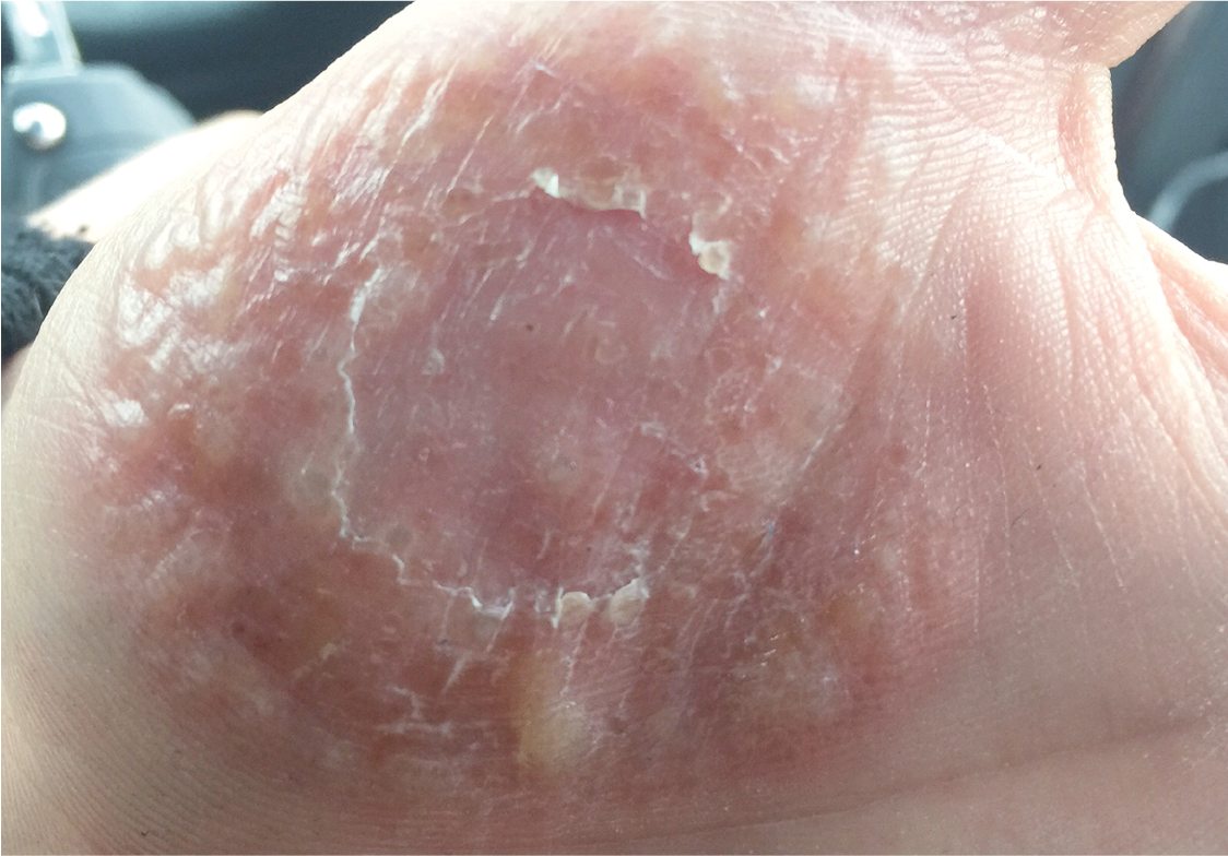
A 20-year-old male reported having bullous lesions on both hands (Fig. 1). However, by the time he presented at the dermatological clinic his bullous lesions had cleared spontaneously, leaving epidermal, squamous lesions. The bullous hand lesions had been waxing and waning for approximately one month and had previously been interpreted as pompholyx. A diagnostic test was obtained.
Fig. 1. The left palm of a patient with bullous lesions. Photograph taken before his first visit to the dermatology department.
Topical treatment with betamethasone had been unsuccessful and had only resulted in partial clearance. The patient had no history of atopic dermatitis. He worked with manual labor in the industry.
What is your diagnosis?
A medical history revealed that the patient kept 2 hedgehogs as pets. Fungal culture on a casein agar plate of a swab from the patient’s hand resulted in microscopic sparse growth of macroconidia and numerous microconidia (Fig. S1). A urease test was weak positive and difficult to evaluate, which raised suspicion, and material was sent to the Centraalbureau voor Schimmelcultures, Fungal Biodiversity Centre Institute of the Royal Netherlands Academy of Arts and Sciences, Utrecht, the Netherlands, for molecular verification.
The PCR sequence obtained was compatible with growth of T. erinacei, a zoonotic dermatophyte commonly known to affect hedgehogs. When first described in 1963, it was classified as a variation of T. mentagrophytes (1). It has later been reclassified as a separate species due to its distinct morphological and physiological characteristics (2). An important difference is the absence or very slow hydrolysis of urea seen in T. erinacei, whereas hydrolysis is rapid in other varieties of T. mentagrophytes (3).
Among humans, T. erinacei have been reported to cause inflammatory and bullous lesions, most often affecting the superficial skin of the hands. Infection with T. erinacei is a rare finding in Europe, and, to our knowledge, this is the first clinical case published in the Scandinavian countries. It has been suggested that findings of T. erinacei as a pathogen in humans could be increasing, as hedgehogs are becoming more popular as domestic pets (4).
The patient was treated with oral terbinafine for 2 weeks, resulting in total clearance. He was asked to take his hedge-hogs to a veterinarian to obtain treatment for the animals and prevent re-infection.
Bullous presentation of dermatophyte infection is a relatively rare clinical presentation and is seen mainly in T. mentagrophytes; however, it constitutes an important differential diagnosis to cutaneous bullous disease, hence the threshold for fungal culture should be low. When tinea manuum is diagnosed, patients should generally be prescribed a systemic antifungal drug. Terbinafine constitutes the first-line treatment and good clinical response is expected and treatment failure is rare. This case illustrates the importance of penetration of domestic animals. Finally, bullous skin lesions might represent Id reaction (autoeczematization) and, in these cases, fungus might be found elsewhere, and not necessarily in the active bullous lesions.
The authors are indebted to Professor Jan Faergemann for his valuable advice in this case.
The authors declare no conflicts of interest.


 Click to show fullsize