


Department of Dermatology, Radboud Institute for Molecular Life Sciences, Radboud University Medical Center, Rene Descartesdreef 1, NL-6525 GL Nijmegen, The Netherlands. *E-mail: Ellen.vandenBogaard@radboudumc.nl
Recent studies have indicated the significance of Th9 cells in a variety of inflammatory and allergy-related diseases (see Kaplan et al. (1)). Th9 cells are a distinct subset of CD4+ T cells present in the skin, which secrete interleukin (IL)-9 (2–4). IL-9 is transiently expressed by skin-tropic and skin-resident Th9 cells to regulate the production of inflammatory cytokines (5). IL-9, like IL-4, is associated with predominantly type 2 immune responses, and aberrant IL-9 expression or signalling in skin is implicated in allergic contact dermatitis (ACD) (6), atopic dermatitis (AD) (7–9), and psoriasis (5, 10). Targeting of IL-9 or its receptor may therefore be an interesting new therapeutic avenue to be explored. Although the IL-9 receptor (IL9R) is expressed by immune cells as well as epithelial cells (11), the majority of research focuses on the Th9/IL-9 axis in immune cells. Recent in vitro studies, however, have shown the regulation of IL9R expression in keratinocytes by IL-4 (12) and increased IL9R expression in basal keratinocytes of psoriatic lesions (10). Furthermore, IL-9 increased IL-8 (CXCL8) and vascular endothelial growth factor (VEGF) secretion by keratinocytes in vitro (6, 7). The effects of IL-9 on human keratinocytes with regard to epidermal proliferation, differentiation and host defence are still poorly understood. This study examined the effects of IL-9 on epidermal morphology, proliferation, differentiation and host defence, and studied cytokine-mediated regulation of IL9R expression on keratinocytes, and investigated the potential additive or synergistic effects by IL-9 in Th2-cytokine mediated epidermal responses.
Human epidermal equivalents (HEEs) and monolayer cultures generated from adult primary human keratinocytes were exposed to human recombinant IL-9 or other pro-inflammatory cytokines, as indicated. Epidermal morphology was studied by histology, and gene and protein expression was analysed by quantitative PCR analysis and immunohistochemistry, respectively. For detailed description, see Appendix S1.
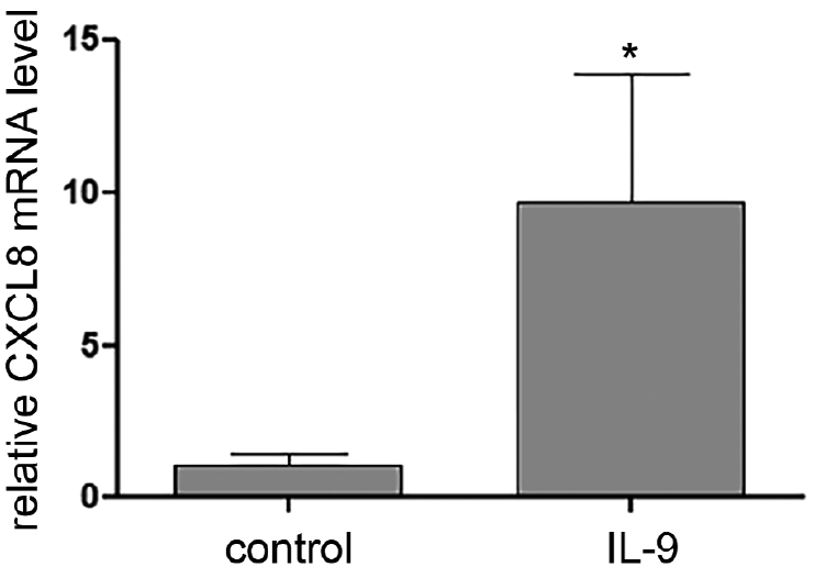
First, a dose range of IL-9 was tested on differentiating submerged keratinocyte monolayer cultures and no effect on cell morphology and viability was observed, even at the highest concentration of 500 ng/ml (data not shown). For further in depth analysis of keratinocyte proliferation and differentiation, HEEs were exposed to 50 ng/ml human recombinant IL-9, being a biologically relevant concentration as shown by the induction of CXCL8 expression (Fig. 1, (10)). After 72 h of IL-9 stimulation, HEEs showed normal epidermal morphology with a fully stratified epidermis and a well-developed stratum corneum (Fig. S1a). The number of proliferating cells, measured with Ki67 staining, and the epidermal thickness was similar to that of control HEEs (Figs S1a and Fig. S2). Next, we analysed the expression of the major epidermal differentiation proteins, keratin 10 (K10), involucrin (IVL), filaggrin (FLG) and loricrin (LOR) (Fig. S1b). For all markers, IL-9 did not alter protein localization or expression levels, nor did it change the expression of epidermal differentiation genes (Fig. S3).
Fig. 1. Interleukin-9 (IL-9) induces epidermal CXCL8 expression. CXCL8 mRNA expression of human epidermal equivalents stimulated with IL-9 (50 ng/ml) for 72 h. Bars represent mean ± standard deviation (SD), n = 3 keratinocyte donors, *p < 0.05.
Also, marker expression for keratinocyte activation or host defence, namely keratin 16 (K16) and SKALP remained unaffected. Human beta defensin 2 (hBD2) is absent in unstimulated HEEs and is also not induced by IL-9 (Fig. S1c). This in contrast to the classical Th2 cytokines, IL-4 and IL-13, which downregulated FLG, LOR and IVL expression (Fig. S4a) and induced K16 and SKALP expression (Fig. S4b).
The suggested role of IL-9 in atopic diseases, which are largely Th2 driven, led us to investigate the interaction of IL-9 with IL-4 and/or IL-13. Co-stimulation with IL-9 did not alter the effect of Th2 cytokines on downregulation of epidermal differentiation proteins (Fig. S5a) or the induction of inflammatory epidermal markers, K16 and SKALP (Figs S5b and Fig. S6).
We hypothesized that the absence of significant effects by IL-9 in our study could be due to low IL9R expression (mean Ct value 34) under the conditions described above. IL9R expression appears to be induced in inflammatory processes, and we therefore stimulated keratinocyte monolayers and HEEs with various cytokine combinations and found interferon gamma (IFN-γ) to be the main inducer of IL9R expression (Fig. 2). We determined the minimal, but effective, IFN-γ exposure time and observed significantly induced IL9R expression after 6 h of IFN-γ stimulation (Fig. S7). Thereafter, HEEs were exposed to IL-9 for 72 h. IFN-γ alone induced the mRNA expression of IVL, and the chemokines CCL5 and CXLC10, yet no additional effect of IL-9 stimulation was observed on these or other genes analysed (Fig. S8A). Similar to previous experiments, IL-9 did not alter epidermal morphology or differentiation protein expression (K10, IVL, LOR and FLG) even under conditions of high IL9R expression due to 6 h IFN-γ pre-stimulation (Fig. S8B).
Fig. 2. IL-9R expression after pro-inflammatory cytokine stimulation. IL9R mRNA expression of submerged keratinocytes stimulated for 48 h with single cytokines or mixes thereof: Th1: IL-1a (30 ng/ml), TNF-α (30 ng/ml), IFN-γ (500 U/ml), Th2: IL-4 (50 ng/ml), IL-13 (50 ng/ml), Th17: IL-17 (30 ng/ml), IL-22 (30 ng/ml). Boxplots represent mean ± standard deviation (SD), n = 3 keratinocyte donors, *p < 0.05.
This study explored the potential effects of IL-9 on keratinocytes and investigated its potential role in multiple biological processes involved in epidermal homeostasis, such as proliferation, differentiation, host defence and inflammatory responses. Even after the induction of IL9R in keratinocytes we did not detect any effect of IL-9 on any of the aforementioned processes, besides the induction of CXCL8 mRNA expression, which has been reported previously (12). Induced expression of this chemokine may potentially aid in the inflammatory process mediated by Th9 cells and cytokines.
Our data indicate that IL9R expression in human keratinocytes is constitutively low, but highly inducible upon inflammatory conditions, and that this regulation is mainly due to IFN-γ. This finding is in line with the upregulated expression of this receptor as found in psoriasis and allergic contact dermatitis (2, 3, 5), where IFN-γ levels prevail and contribute to disease pathogenesis. In contrast, Hong et al. (12) found IL9R to be upregulated by IL-4. We did not detect Th2-cytokine mediated induction of IL9R mRNA expression in HEEs (Fig. 1A) or monolayer cultures (data not shown). The differences in cell source or post-transcriptional regulation may explain this discrepancy, since Hong et al. studied IL9R protein expression in foreskin keratinocytes.
The limitation of the current study is that we examined a selection of genes. Genome-wide transcriptomic analysis may reveal additional IL-9 responsive genes in epidermal keratinocytes, other than CXCL8 reported here, but this is outside the scope of the current study. Based on our data we conclude that IL-9 does not affect important keratinocyte functions, such as proliferation or transcription of major differentiation-related genes involved in skin barrier function.
The authors declare no conflicts of interest.


 Click to show fullsize
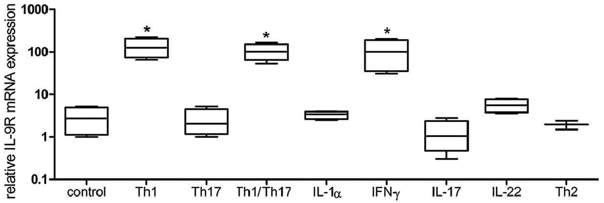 Click to show fullsize