


Department of Pathophysiology and Transplantation, Università degli Studi di Milano, IRCCS Foundation, Cà Granda Ospedale Maggiore Policlinico, via Pace n.9, IT-20122 Milan, Italy. E-mail: stefano.veraldi@unimi.it
A 46-year-old man was referred to our department by his general practitioner with a clinical diagnosis of melanoma on the fifth left toe. The patient reported that he was in good general health and not on any systemic drug therapy. The slightly painful lesion had appeared approximately 6 weeks earlier, during a trip to Fortaleza (Brazil).
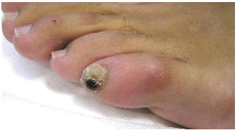
Dermatological examination revealed a pigmented, slightly infiltrated lesion, approximately oval in shape, 0.8 × 0.5 cm, brownish-black in colour, with poorly defined borders (Fig. 1). Dermoscopy of the lesion is shown in Fig. 2.
Fig. 1. Pigmented, slightly infiltrated lesion, oval in shape, brownish-black in colour, with poorly defined borders.
Fig. 2. Grey-yellowish round lesion with a central black pore corresponding to the posterior portion of the abdomen of the flea. A haemorrhagic ring surrounds the whole lesion.
What is your diagnosis? See next page for answer.
Acta Derm Venereol 2017
Diagnosis: Tungiasis
Tungiasis is an infestation caused by penetration into the skin by the gravid female of the flea Tunga penetrans Linnaeus 1758 (Insecta, Siphonaptera: Tungidae). Tungiasis is characterized clinically by one or more papular or nodular lesions, grey or yellowish in colour, with a small central brown-black opening corresponding to the posterior portion of the abdomen of the flea. It is usually located on the feet (toes, peri- and subungual folds, interdigital folds, sole and heel). Diagnosis is usually easy. However, less common clinical varieties of tungiasis have been described: plantar wart-like lesions, as well as crusted, bullous, pustular and ulcerative lesions (1).
Dermoscopy is useful in the diagnosis of infections and infestations of the skin (2, 3), including tungiasis (2–14). The dermoscopic picture of tungiasis is characterized by a whitish to light brown nodule, with a peripheral pigmented ring surrounding a black central pore, corresponding to the posterior portion of the abdomen of the flea (2–4, 7, 10, 12, 13). In addition, grey-blue blotches (2, 3, 6, 10) and whitish oval structures linked together to form chain-like structures (“whitish chains”) (3, 9) may be seen, corresponding to the eggs in the abdomen of the flea. Clusters of eggs may also be observed as jelly-like bags (3, 7, 13). Furthermore, a “radial crown” has been described (10).
The lesion was removed successfully by curettage (Fig. 3). Parts of the body of the flea and some eggs were observed. A 3-month follow-up was negative.
Fig. 3. The lesion was successfully removed by curettage.


 Click to show fullsize
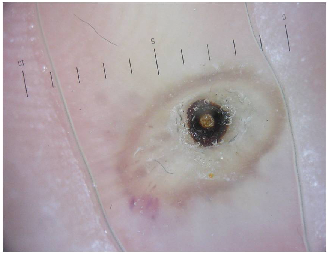 Click to show fullsize
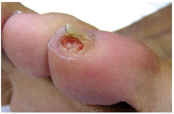 Click to show fullsize