


Departments of 1Dermatology and 3Pathology, 4Institut Mar d’Investigacions Mèdiques (IMIM), Universitat Autònoma de Barcelona, Hospital del Mar-Parc de Salut Mar, Passeig Marítim 25–29, ES-08003 Barcelona, and 2Department of Dermatology, Hospital Josep Trueta, Girona, Spain. *E-mail: 93329@parcdesalutmar.cat
Accepted Apr 3, 2017; Epub ahead of print Apr 4, 2017
Peripheral blood eosinophilia has been reported to occur in a wide range of haematological malignancies including primary cutaneous lymphomas. The concurrence of lymphomatoid papulosis (LyP) with peripheral blood eosinophilia seems to be an uncommon phenomenon. Atypical lymphocytes in LyP display the phenotype of activated T-helper cells, consistently express CD30 antigen, have a Th2 cytokine profile, and secrete eosinophil-stimulating cytokines. Cases of LyP have been reported in association with myeloproliferative hypereosinophilic syndrome (M-HES) presenting the Fip1-like 1/platelet-derived growth factor receptor-α (FIP1L1-PDGFRA) fusion gene. In such cases, imatinib treatment may lead to a complete and persistent resolution of LyP lesions.
A 25-year-old man was referred to our Department in November 2011 for evaluation of a 3-year history of recurrent crops of self-healing erythematous papules and nodules with central ulceration on the lower extremities, oral mucosa and face. Biopsy had established the diagnosis of LyP. The patient reported periodical appearance of lesions that regressed spontaneously leaving residual scars. His past medical history revealed childhood-onset asthma, allergic conjunctivitis and rhinitis. Peripheral eosinophilia of 1,200/mm3 had also been detected and was related to the allergic symptoms along with progressive cough and discrete dyspnoea.
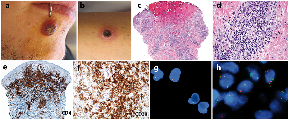
Physical examination disclosed a firm, well-demarcated, painless, ulcerated nodule, 1.2 cm in diameter on the right aspect of the chin. Several erythematous papules and residual scars on the lower extremities were also observed. A solitary nodule with a central crust was noted on the left leg (Fig. 1a, b). No enlarged lymph nodes were detected.
Fig. 1. Clinical and histochemical characteristics. (a) Ulcerated inflammatory nodule on the right side of the patient’s chin. (b) Necrotic erythematous infiltrated plaque on the leg. (c) Histopathology revealing a wedge-shaped dense inflammatory infiltrate extending from papillary to reticular dermis (haematoxylin and eosin staining (H&E); original magnification ×40). (d) Atypical, enlarged lymphocytes admixed with smaller lymphocytes, neutrophils and eosinophils (H&E ×400). (e) The infiltrate showed positive CD4 immunoreactivity (CD4 stain; ×40). (f) Atypical, large lymphocytes corresponded with CD30+ cells (CD30 stain; ×400). (g) Detection of the (4q12) deletion by interphase fluorescence in situ hybridization (FISH) in peripheral blood cells. Loss of red signal indicating fusion of FIP1L1-PDGFRA. (h) FIP1L1-PDGFRA fusion gene was not detected in T cells from cutaneous LyP lesion by FISH.
Two 4-mm punch biopsies from different lesions disclosed a dense, dermal, wedge-shaped infiltrate with clusters of large atypical lymphoid cells admixed with small lymphocytes, histiocytes, neutrophils and eosinophils (Fig. 1c–f). Neoplastic cells were CD30+ and expressed mature T helper cell markers (CD3, CD2, CD5 and CD7, CD4). No expression of ALK, c-KIT or CD56 was detected. Neoplastic cells were also MUM-1+ and 60% of cells expressed Ki-67 antigen. In both biopsy specimens, an identical clonal rearrangement of the T-cell receptor (TCR) γ and β chains was detected. No T-cell clonality was detected in peripheral blood samples.
Laboratory tests disclosed massive peripheral eosinophilia (6,120/mm3), normal IgE levels, elevated tryptase (15.5 ng/ml) (NV < 13.5) and vitamin B12 serum levels (>2,000 ng/ml) (NVs: 197–866). Stools for ova and parasites were negative. Body computed tomography (CT) showed bronchial dilatation with peribronchial thickening and confluent centrolobulillar nodules forming a ground glass pattern. Electrocardiogram, echocardiogram and ophthalmological examination showed no abnormalities.
A bone marrow biopsy was obtained showing hypercellular marked eosinophilia, atypical spindle-shaped mast cells and increased reticulin fibrosis. Interstitial deletion of the 4q12 chromosome band comprising LNX, CHIC2 and PDGFRA genes leading to a FIP1L1-PDGFRA rearrangement was detected by fluorescence in situ hybridization (FISH) in peripheral blood cells (Fig. 1g). The deletion was present in eosinophils (46%), mast cells (100%) and granulocytes (45%) analysed from peripheral blood isolated populations. This led to the diagnosis of hypereosinophilic syndrome secondary to FIP1L1-PDGFRA myeloid neoplasm. The presence of FIP1L1-PDGFRA fusion gene was analysed by FISH in skin biopsy specimens of LyP. No individual cells were found to bear the translocation (Fig. 1h).
Treatment with imatinib mesilate (100 mg/day) was prescribed in February 2012. Regression of the cutaneous lesions and a rapid normalization of the eosinophil count were observed. After 8 months of treatment, a sustained response of both haematological profile and LyP lesions was achieved and dosage was progressively tapered. After 58 months of treatment, the patient remains asymptomatic on a maintenance dosage of 100 mg/week (Fig. S1).
Characteristically, LyP is manifested by recurrent development of papulo-nodular lesions that undergo spontaneous regression after weeks or months. Many different therapeutic options have been proposed; nevertheless, after treatment discontinuation, relapses tend to occur (1) and no therapy has proven to alter the course of the disease nor prevent LyP-associated lymphomas.
Since the original description, 11 cases of LyP associated with idiopathic HES have been reported (2–8). The associated HES corresponded to M-HES and specifically to patients presenting the FIP1L1-PDGFRA fusion gene, the most well-characterized molecular defect leading to hypereosinophilia (9). This subset of HES is characterized by splenomegaly, elevated serum B12 and tryptase levels, myeloproliferation and tissue fibrosis. Patients usually show a dramatic response to imatinib, which targets these genes (10).
The mechanisms implicated in the concurrent development of M-HES and LyP remain obscure. Clonal T cells bearing the FIP1L1-PDGFRA rearrangement have been identified in some patients with PDGFRA-associated HES, moreover, cases associating M-HES and T-cell lymphoblastic lymphoma in which both disorders carried the same FIP1L1-PDGFRA rearrangement have been reported (11, 12). Conversely, a non-LyP CD30+ clonal T-cell proliferation associated with non-FIP1L1-PDGFRA HES has been described recently, supporting the role of HES in the development of a clonal T-cell proliferations (13). A “multilineage clonal population” has also been postulated as the most likely explanation for the M-HES-LyP association (14). However, in our case we have been unable to demonstrate the presence of the specific translocation in cutaneous infiltrates of LyP lesions. The possibility that the neoplastic population bearing the FIP1L1-PDGFRA gene fusion could be beyond the threshold of FISH detection, although improbable, cannot be disregarded. Therefore, the possibility that independent genetic events may engender this association cannot be excluded.
Since 2003, imatinib was prescribed in 7 cases of M-HES associated with LyP (Table SI). In 4 of these, the FIP1L1-PDGFRA fusion gene was demonstrated. A rapid regression of the cutaneous lesions was noted and a persistent, complete clinical, haematological and molecular remission was achieved (4, 6). No recurrences of LyP have been observed after variable follow-up periods. In our case, no new lesions have developed after a 5-year follow-up period.
The mechanisms implied in this constant response have not been elucidated. This parallel effect in both LyP and M-HES has not been observed in cases previously treated with regimes not including imatinib (2, 3). These observations could suggest that tyrosine kinase-mediated mechanisms play a role in the development of LyP. Furthermore, the possibility that this therapeutic effect could be a phenomena not restricted to the subset of LyP patients associated with M-HES has not been evaluated.
The authors declare no conflicts of interest.


 Click to show fullsize