


1CHRU Brest, Department of Pathology, 2European University of Brittany, and 3INSERM, U1078, Brest, France
BRAF and NRAS genetic analyses are time-consuming and can delay treatment choices in patients with metastatic melanomas presenting with acute deterioration. We compared the rapid, real-time, fully automated molecular diagnosis platform Idylla™ with next-generation sequencing (NGS) and immunohistochemistry for detection of BRAF and NRAS mutations in 36 patients with metastatic melanomas. The Idylla™ NRAS-BRAF-EGFRS492R mutation assay (110 min per sample) detected BRAF and NRAS mutations in 15 and 17 samples, respectively. One NRAS mutation was different between NGS and Idylla™ (NRASG13C vs. NRASG12A/D). Four samples were BRAF and NRAS wild-type. The global concordance between NGS and Idylla™ assays was 97.2% (35/36 cases). Immunohistochemistry was positive only in 9/9 BRAFV600E- and 6/6 NRASQ61R-mutated samples with VE1 and SP174 antibodies, respectively. The Idylla™ platform is a valuable rapid molecular diagnosis tool to reduce the delay in BRAF and NRAS analyses-related treatment choices for patients with metastatic melanoma presenting with acute deterioration.
Key words: melanoma; BRAF; NRAS; immunohistochemistry; Idylla; next-generation sequencing
Accepted Jun 28, 2017; Epub ahead of print Jun 29, 2017
Acta Derm Venereol 2017; 97: xx–xx.
Corr: Arnaud Uguen, Department of Pathology, University Hospital Morvan, 5, Avenue Foch, FR-29609 Brest, France. E-mail: arnaud.uguen@chu-brest.fr
Melanoma is a frequent and aggressive skin cancer with a high rate of mortality at the metastatic stage (1–3). The management of patients with metastatic melanoma has improved recently with immunotherapy and molecular-targeted therapy (4–7). Current molecular-targeted therapies consist mainly of BRAF and MEK inhibitors used in patients with BRAF-mutated meta-static melanomas, which represent approximately 50% of melanomas (4, 5, 8). In contrast, there is currently no approved targeted therapy to inhibit NRAS mutant proteins, which are detected in approximately 15% of melanomas, and patients with NRAS-mutated metastatic melanomas are treated with immunotherapy (6–8). Thus, the determination of BRAF and NRAS mutation status in melanoma samples is now a major criterion for treatment choices (9, 10).
The method of detection of BRAF and NRAS mutation should be accurate, highly sensitive to detect low mutant allele frequency, and fast enough to provide useful information allowing rapid treatment choice in patients with metastatic melanomas. The vast majority of samples analysed for mutation testing are formalin-fixed and paraffin-embedded (FFPE) melanoma samples from pathology centres. In addition to the DNA-based, PCR-based, and sequencing methods, such as next-generation sequencing (NGS), mutation-specific immunohistochemistry (IHC) has emerged recently as an efficient and more rapid tool in comparison with sequencing to detect BRAFV600E and NRASQ61R mutant proteins in FFPE melanoma samples using the clones VE1 and SP174, respectively (11–15). Although BRAFV600E and NRASQ61R mutations represent approximately 90% of BRAF mutations and 40% of NRAS mutations in melanoma, respectively, other BRAFV600 and NRAS mutations in codons 12, 13 and 61 are also relevant for treatment choice, but are not detected with clones VE1 and SP174. Thus, the detection of different BRAFV600 and NRAS codons 12, 13 and 61 mutations still requires expensive molecular biology equipment and a dedicated laboratory with staff highly skilled in molecular methods, in order to provide a clinically relevant BRAF and NRAS mutation status to the clinicians. The molecular testing process of a tumour sample can last several days to weeks depending on institutions, which can delay the start of targeted therapy or immunotherapy in patients with metastatic melanomas (16).
Recently, a fully automated real-time PCR Idylla™ platform has been developed to provide rapid detection (less than 2 h) of BRAF mutation in FFPE melanoma samples (17–20). It has shown excellent performances in comparison with other DNA-based methods, including NGS, but also with BRAFV600E mutation-specific IHC. However, until recently, this platform did not provide any information about NRAS mutation status, which still required additional sequencing analyses with an alternative method in melanomas.
The aim of the present study is to compare the new Idylla™ fully-automated platform with NGS and mutation-specific IHC to detect BRAF and NRAS mutations in melanoma samples. This is the first report of a rapid, fully automated real-time PCR method enabling the detection of BRAF and NRAS mutations in the same assay in melanomas.
Thirty-six FFPE melanoma samples were collected from patients selected from the cases analysed by the Brest University Hospital cancer molecular genetics platform in 2015 and 2016. BRAF and NRAS analyses were conducted as part of the diagnostic work-up for the therapeutic management of patients with advanced stages of melanoma according the recommendations of the French National Cancer Institute. The samples were enriched in BRAF- and NRAS-mutated samples according to initial NGS results and were selected in order to evaluate the performance of the IdyllaTM platform with different mutations. All samples were included in a registered tumour tissue collection, and the present study was conducted in compliance with the principles of the Declaration of Helsinki, following approval by our institutional review board (CHRU Brest, CPP number DC – 2008 – 214).
The proportion of tumour cells in each sample was established by a pathologist on a dedicated 3 µm-thick tissue slide stained with haematoxylin-eosin-saffron (HES). Serial 10 µm and 5 µm unstained tissue sections were produced for molecular and IHC analyses, respectively. The tumour zones were macroscopically circled to allow macrodissection of tumour tissue for genetic analyses (NGS and IdyllaTM analyses) whenever possible (i.e. clear delimitation between tumour and non-tumour adjacent tissue).
The monoclonal antibodies N-Ras (Q61R) (clone SP174, Spring Bioscience, Pleasanton, CA, USA) and BRAF V600E (clone VE1, Spring Bioscience) were used at a dilution of 1:100. IHC was performed on Ventana Benchmark XT® automated slide preparation system (Roche Diagnostics, Meylan, France) using ultraView Universal Alkaline Phosphatase Red Detection Kit (Roche Diagnostics), as reported previously (15, 21). UltraView® Red detection kit was used through Ventana staining procedure that included pretreatment with cell conditioner 1 (pH 8) for 60 min, followed by incubation with diluted antibody at 37°C for 32 min. Antibody incubation was followed by standard signal amplification with the Ventana amplifier kit and ultra-Wash. Slides were counterstained with 1 drop of haematoxylin for 12 min and 1 drop of bluing reagent for 4 min. Subsequently, the slides were removed from the immunostainer, washed in water with dishwashing detergent, and mounted.
Immunostaining was interpreted by a single pathologist with-out knowledge of the molecular status. Staining was considered positive when it was cytoplasmic and moderate to strong, clearly different from the background. It was considered negative when no or only faint or nuclear labelling was noted.
NGS analyses were performed as reported previously (15). Maxwell 16 CE-IVD system (Promega Corporation, Fitchburg, WI, USA) combined with the Maxwell® 16 FFPE Tissue LEV DNA Purification Kit (Promega Corporation) was used to isolate DNA from 3 series of 10-µm sections of dissected tissue blocks. DNA was eluted with 100 µl water provided by the manufacturer. DNA libraries were produced using custom Ion AmpliSeq™ Panel (Life Technologies, Villebon sur Yvette, France) according to the manufacturer’s instructions. After libraries quantification by qPCR (Ion Library Quantitation kit, Life Technologies) and Roche 480 Lightcycler Real-Time PCR), 15 bar-coded (Ion Xpress Barcodes adapters kit, Life Technologies) tumour DNA libraries were sequenced simultaneously on a 316 chip in the Personal Genome Machine (PGM) system (Ion Torrent, Life Technologies). Torrent suite software v4.4.0 was used for signal processing, run quality report and Fastq files generation. BRAF and NRAS sequences were then analysed through the SeqNext software v4.1.2 (JSI Medical Systems GmbH, Ettenheim, Germany). Nucleotide numbering was carried out in accordance with Human Genome Variation Society (HGVS) recommendations (www.hgvs.org/mutnomen). The reference sequences NM_004333.4 for BRAF gene and NM_002524.4 for NRAS gene were used for cDNA-based numbering, i.e. the A of the ATG translational initiation codon was ascribed as +1.
The Idylla™ platform (Biocartis, Mechelen, Belgium) is a fully cartridge-based automated platform and uses microfluidics processing with all reagents on-board. In our study we used the Idylla™ NRAS-BRAF-EGFRS492R Mutation Assay cartridge (Biocartis) initially designed for colorectal carcinomas. The 36 melanoma samples were assessed for the detection of BRAFV600E (c.1799T>A; p.Val600Glu and c.1799_1800TG>AA; p.Val600Glu i.e. BRAFV600E2 variant), BRAFV600D (c.1799_1800TG>AC; p.Val600Asp), BRAFV600K (c.1798_1799GT>AA; p.Val600Lys), BRAFV600R (c.1798_1799GT>AG; p.Val600Arg), NRASG12C (c.34G>T; p.Gly12Cys), NRASG12S (c.34G>A; p.Gly12Ser), NRASG12D (c.35G>A; p.Gly12Asp), NRASG12A (c.35G>C; p.Gly12Ala), NRASG12V (c.35G>T; p.Gly12Val), NRASG13D (c.38G>A; p.Gly13Asp), NRASG13V (c.38G>T; p.Gly13Val), NRASG13R (c.37G>C; p.Gly13Arg), NRASA59T (c.175G>A; p.Ala59Thr), NRASQ61R (c.182A>G, p.Gln61Arg), NRASQ61K (c.181C>A; p.Gln61Lys), NRASQ61L (c.182A>T; pGln61Leu), NRASQ61H (c.183A>C or c.183A>T; p.Gln61His), NRASK117N (c.351G>C or c.351G>T; p.Lys117Asn), NRASA146T (c.436G>A; p.Ala146Thr), NRASA146V (c.437C>T; p.Ala146Val) and EGFRS492R (c.1476C>A or c.1474A>C; p.Ser492Arg) mutations. For each sample, 1–3 slides were used to obtain a total macrodissected area of 5–30 mm2 FFPE tumour tissue according to the manufacturer’s instructions and then transferred to a wetted (nuclease-free water) filter paper. A second wetted filter paper was then added on top of the FFPE material and the sample with the 2 wetted filter papers was finally placed on the lysis pad in the Idylla™ NRAS-BRAF-EGFRS492R Mutation Assay cartridge and inserted in the instrument. Inside the cartridge, the sample was homogenized and cells lysed using a combination of high-intensity focused ultrasound, enzymatic/chemical digestion and heat. The nucleic acids were liberated and ready for subsequent PCR amplification. The PCR was real-time and used a fluorophore-based detection system. After a 112-min run, all steps were performed automatically inside the cartridge and final reports were directly available on the system after an automatic on-board post-PCR curve analysis. BRAFV600E, BRAFV600E2 and BRAFV600D mutations were detected by the system as “V600E/D Mutation”, whereas BRAFV600K and BRAFV600R mutations were detected as “V600K/R Mutation”. NRASG12C, NRASG12S, NRASG12D, NRASG13D, NRASA59T, NRASQ61R, NRASQ61K, NRASQ61L, NRASQ61H, NRASK117N and EGFRS492R mutations were individually detected by the system, whereas NRASG12A and NRASG12V mutations were detected as “G12A/V Mutation”, NRASG13V and NRASG13R mutations as “G13V/R Mutation” and NRASA146T and NRASA146V mutations as “A146T/V Mutation”. Total BRAF, NRAS and EFGR acted as sample processing controls (data not shown by the system).
Of the 36 cases included in this study, 12 were primary melanomas and 24 were metastases (see Table I for details). The mean percentage of tumour cells on whole slides was 62% (20–95%) and tissue macrodissection permitted an increase in the mean percentage of cells used for molecular analyses to 76% (50–95%). Fifteen samples were BRAF-mutated (9 BRAFV600E mutations including 2 BRAFV600E2 variants, 4 BRAFV600K and 2 BRAFV600R mutations) and 17 samples were NRAS-mutated (6 NRASQ61R, 4 NRASQ61L, 3 NRASQ61K, 2 NRASQ61H, 1 NRASG13D, and 1 NRASG13C mutations) according to NGS analyses.
Table I. Detailed clinical and pathological data with molecular and immunohistochemistry (IHC) results of the 36 melanoma samples
The 9 BRAFV600E-mutated samples were positive with VE1 IHC with homogeneous strong-to-moderate staining (Fig. 1). SP174 stained homogeneously the 6 NRASQ61R-mutated melanomas (Fig. 2). The 21 samples being neither BRAFV600E- nor NRASQ61R-mutated were negative with VE1 and SP174 IHC.
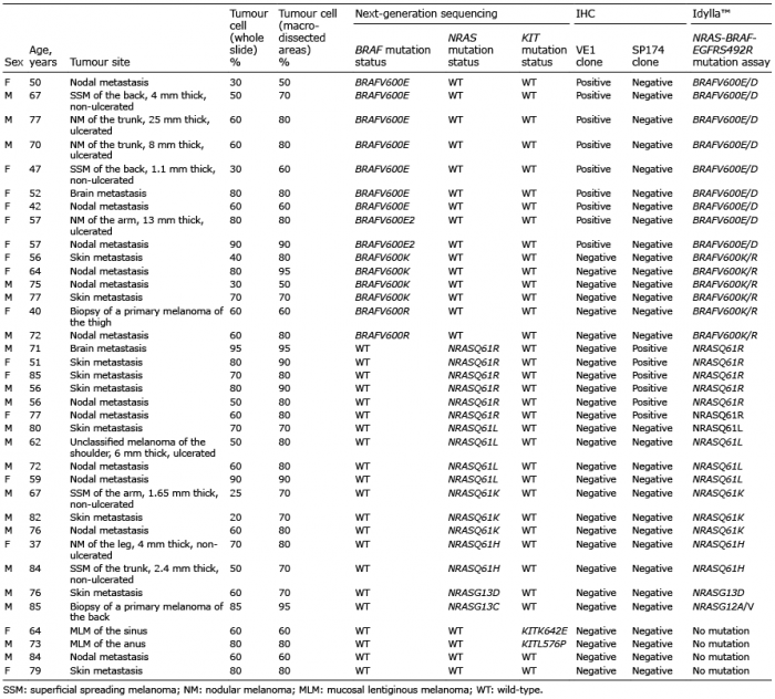
Fig. 1. Example of a BRAFV600E-mutated primary melanoma in a 67-year-old man. (a) The sample was found to contain approximately 50% of tumour cells (haematoxylin-eosin-saffron staining, ×2.5 magnification). (b) VE1 anti-BRAFV600E immunohistochemistry (IHC). The sample was strongly positive (Red revelation, haematoxylin counter-coloration, ×2.5 magnification). (c) SP174 anti-NRASQ61R IHC. The sample was considered as negative (Red revelation, haematoxylin counter-coloration, ×2.5 magnification). (d) Idylla™ NRAS-BRAF-EGFRS492R mutation assay. The sample was concluded as having a “BRAFV600E/D mutation” (screenshot of the Idylla™ automated report).
Fig. 2. Example of a NRASQ61R-mutated mutated lymph node metastasis in a 77-year-old woman. (a) The sample was found to contain approximately 60% of tumour cells. haematoxylin-eosin-saffron staining, ×1.5 magnification). (b) VE1 anti-BRAFV600E immunohistochemistry (IHC). The sample was considered as negative (Red revelation, haematoxylin counter-coloration, ×1.5 magnification). (c) SP174 anti-NRASQ61R IHC. The sample was strongly positive (Red revelation, haematoxylin counter-coloration, ×1.5 magnification). (d) Idylla™ NRAS-BRAF-EGFRS492R mutation assay. The sample was concluded as having a “NRASQ61R mutation” (screenshot of the Idylla™ automated report).
Using the Idylla™ platform, the 9 BRAFV600E-mutated samples were concluded having a “BRAFV600E/D Mutation” whereas the 4 BRAFV600K-mutated and the 2 BRAFV600R-mutated samples were concluded having a “BRAFV600K/R Mutation”. The fully automated platform also rightly identified the NRAS mutations in the 6 NRASQ61R-, 4 NRASQ61L-, 3 NRASQ61K-, 2 NRASQ61H- and 1 NRASG13D-mutated samples (see Figs 1 and 2). A single sample presented a different NRAS mutation between the results of NGS and Idylla™ analyses with a NRASG13C (c.37G>T; p.Gly13Cys) mutation according to NGS and a “NRASG12A/D mutation” detected by the Idylla™ platform (Fig. 3). A third molecular SNaPshot method also concluded in a NRASG13C mutation in this discrepant sample. The global concordance between NGS and Idylla™ was 97.2% (35/36 cases).
Fig. 3. Primary melanoma of the back in an 85-year-old man with discrepant results between next-generation sequencing and Idylla™ assays. (a) The sample was found to contain approximately 85% of tumour cells (haematoxylin-eosin-saffron staining, ×4 magnification). (b) Next-generation sequencing. Identification of a c.37G>T (p.G13C) mutation in NRAS codon 13 (65.26%). (c) Idylla™ NRAS-BRAF-EGFRS492R mutation assay. The sample was concluded as having a “NRASG12A/V mutation” (screenshot of the Idylla™ automated report).
The determination of BRAF and NRAS molecular status has now become mandatory to treat patients with metastatic melanomas (9, 10). This determination often requires expensive equipment, such as NGS solutions, and staff highly skilled in molecular methods. Thus, the molecular tests are restricted mainly to specialized laboratories. There could be a long time between the initiation of analysis and provision of the written report, which could delay the treatment of patients (16). Different methods can be used to provide a more rapid molecular diagnosis in an intent-to-treat strategy.
In the present work and in previous studies, mutation-specific IHC showed good performances compared with molecular methods in melanoma samples (11–15, 21–23). Because IHC is easy to automate and is a widely distributed technique in comparison with molecular methods, it could be an interesting ancillary tool to provide a fast mutation status analysing FFPE melanoma samples in pathology laboratories. Nevertheless, highly sensitive and specific antibodies are, to date, restricted to the most frequent BRAFV600E (clone VE1) and NRASQ61R (clone SP174) mutant proteins in melanomas. New anti-NRASQ61L antibody (clone 26193, NewEast Biosciences) was reported recently as another ancillary tool in melanoma, but it appeared less sensitive than VE1 and SP174 IHC (24). To date, IHC does not allow the detection of other frequent or rare mutations in melanoma, such as other BRAFV600 variants that also respond to anti-BRAF/MEK targeted therapies.
Other ancillary molecular methods are being developed to provide a fast mutation status. Among these techniques, the fully automated Idylla™ platform has been reported recently to have good performance in the detection of BRAF mutation in comparison with high-resolution melting, real-time allele-specific amplification, NGS and IHC in melanoma samples using a Idylla™ BRAF mutation test cartridge (Biocartis) (17–20). This fully automated platform required less than 2 min for FFPE sample preparation and provided a BRAF mutation status in approximately 90 min. It also had the major advantage that it did not require any molecular biology dedicated space for DNA processing, because all the processes, including DNA extraction and analysis, took place inside sealed cartridges, thus preventing any contamination.
In our study, we first used the Idylla™ NRAS-BRAF-EGFRS492R Mutation Assay cartridge initially designed for colorectal carcinomas to detect NRAS and BRAF mutations in melanomas. Demonstrating good performance with this new test in colorectal carcinoma samples, Colling et al. (25) reported a limit of detection inferior to 5% . In the present study, macrodissection of tumour areas by a pathologist allowed the required tumour cells to be obtained in percentages greater than 50% for the 36 assays, with no poor cell samples. Idylla™ assays enabled the accurate detection of every BRAF- and NRAS-mutated melanoma sample, providing a mutation status in under 2 h per sample. Final reports were directly available on the system, providing immediately usable information for treatment choices.
Interestingly, one case with a NRASG13C mutation, which was not included in the set of mutations detected by the Idylla™ NRAS-BRAF-EGFRS492R Mutation Assay cartridge, was detected as a “NRASG12A/D mutation”. Thus, we hypothesized that the primers included in the cartridge for real-time PCR to detect single nucleotide polymorphisms were not absolutely specific for NRASG12A (c.35G>C) and NRASG12V (c.35G>T) mutations, but also permitted detection of the NRASG13C (c.37G>T) mutation. None of the 4 BRAF and NRAS wild-type samples resulted in false-positive results. Facing with our single false result of Idylla™ analyses in our set of 36 samples, we hypothesized that additional methods, such as NGS, must be performed after a first-step fast Idylla™-based mutation diagnosis to precisely type the mutation detected by the Idylla™ platform. Moreover, the Idylla™ NRAS-BRAF-EGFRS492R Mutation Assay cartridge was not designed to detect every rare mutation in BRAF and in NRAS, but also in other oncogenes, such as KIT. In this manner, ancillary molecular analyses would also be required to detect these rare mutations, which are also potentially relevant for treatment decisions. Nevertheless, in our experience, the Idylla™ NRAS-BRAF-EGFRS492R Mutation Assay cartridge was designed to detect more than 95% of the BRAF and NRAS mutations encountered in melanoma samples in our daily practice (data not shown).
In this study, the fully automated Idylla™ platform was found to be a rapid, easy to use diagnostic tool, which was able to provide a BRAF and NRAS mutation status in less than 2 h (110 min) on the basis of FFPE melanoma samples. This test, which is now certified CE-IVD (for in vitro diagnosis) as the Idylla™ NRAS-BRAF mutation test, showed good performance in comparison with NGS and IHC, and could represent an interesting first-step molecular diagnostic tool to reduce delay in treatment choices due to mutation testing in patients with metastatic melanoma presenting with acute deterioration.
The authors would like to acknowledge Sandrine Duigou, Marina Pochic and Véronique Fainsin (CHRU Brest, Department of Pathology) and David Dejans (Biocartis) for their technical assistance, the Local tumor tissue biobank BB-0033-00037 (“CRB Santé/Tumorothèque de Brest”) for their collaboration in this work and “Omnium Group” for support. The authors would also like to thank the molecular geneticists of the Brest University Hospital cancer molecular genetics platform for their daily collaboration in diagnosis practice.
The authors declare no conflicts of interest.


 Click to show fullsize
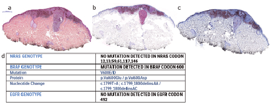 Click to show fullsize
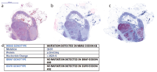 Click to show fullsize
 Click to show fullsize