


Department of Dermatology, University Hospital, Jena, Germany
Cutaneous pseudolymphoma (CPL) is a reactive polyclonal T- or B-cell lymphoproliferative process. CPL may appear as localized or disseminated skin lesions. While most cases of CPL are idiopathic, they may also occur as a response to, for example, contact dermatitis, arthropod reactions, and bacterial infections. CPL can be classified based on its clinical features, but all variants have similar histopathological patterns of either predominantly B-cell infiltrates, T-cell infiltrates, or mixed T/B-cell infiltrates. The prognosis of CPL is good, but the underlying disease process should be taken into account. If an antigenic stimulus is identified, it should be removed. In patients with idiopathic CPL, a close follow-up control strategy should be adopted. The aim of this systematic review is to summarize all reported treatments for CPL. The review was based on articles from the PubMed database, using the query “skin pseudolymphoma treatment”, English and German, about “human” subjects, and published between 1990 and 2015 documenting adequate treatment and/or aetiology. Mainly individual case reports and small case series were found. Treatment options include topical and intralesional agents, systemic agents, and physical modalities. The final part of the review proposes a treatment algorithm for CPL according to each aetiology, based on the literature of the last 25 years. Future research should focus on randomized controlled trials and studies on long-term outcomes, which were not identified in the current review.
Key words: cutaneous pseudolymphoma; cutaneous lymphoid hyperplasia; treatment; lymphadenosis benigna cutis; lymphocytoma cutis.
Accepted Nov 13, 2017; Epub ahead of print Nov 14, 2017
Acta Derm Venereol 2018; 98: XX–XX.
Corr: Diana Miguel, Department of Dermatology, University Hospital, Erfurter Str. 35, DE-07740 Jena, Germany. E-mail: Diana.Miguel@med.uni-jena.de
Cutaneous pseudolymphoma (CPL) is a reactive polyclonal T- or B-cell lymphoproliferative process that develops in reaction to diverse known and un-known stimuli, which can be localized or disseminated in the skin. Pseudolymphomas are also denominated cutaneous lymphoid hyperplasia (CLH), which better describes their benign clinical course. Sarcomatosis cutis, lymphocytoma cutis, lymphadenosis benigna cutis, pseudolymphoma of Spiegler and Fendt, and actinic reticuloid are other accepted terminologies to describe CPL (1, 2). Most cases of CPL are idiopathic. Reactive responses that can result in CPL include responses to contact dermatitis, lichenoid pigmented purpuric dermatosis, lichen sclerosus et atrophicus, the inflammatory stage of morphea, secondary syphilis, lupus panniculitis, arthropod reactions, nodular scabies, viral infections (orf, milker’s nodule, herpes simplex/zoster, and molluscum contagiosum), tattoo dye, vaccinations, trauma, jewellery for pierced ears, such as gold, acupuncture, infections with Borrelia burgdorferi or leishmaniasis, and drug reactions (3). Drugs associated with the development of CPL include anticonvulsants, antipsychotics, antihypertensives, cytotoxics, antirheumatics, antibiotics, anxiolytics, antihistamines, antiarrythmics, sex steroids, lipid-lowering agents, and more recently, anti-tumour necrosis factor (TNF)-α agents, tocilizumab, and cyclosporine (1, 4–6). Although they appear more often in the skin, pseudolymphomas have also been described in other organs, such as the eye, tongue, parotid gland, larynx, gastrointestinal tract, lung, kidney, and breast (4).
In order to diagnose CPL it is important to understand that neither clinical nor histological features alone allow correct classification as lymphoma or pseudolymphoma. Only a combination of clinical signs, histological features and the course of the disease can result in the correct diagnosis. Sometimes a careful drug history, serological tests and patch tests may help to distinguish CPL from lymphoma.
Nevertheless, and because progression of CPL to malignant lymphoma can occur, perhaps induced by persistent antigenic stimulation, regular follow-up of the patient is mandatory (7). However, progression to cutaneous lymphoma has been observed in only a minority of cases.
Clinically, CPL may present as papules, infiltrated plaques and nodules (see examples in Figs 1 and 2), less frequently as persistent erythema or exfoliative erythroderma. Even though there is no single clinical feature that proves a malignant or benign lesion, multiple nodules or plaques substantiate the suspicion of a malignant lymphoma. Lymphadenopathy is also more suggestive for lymphoma (8). Nonetheless, the mixed type of B- and T-cell CPL may also show lymphadenopathy.
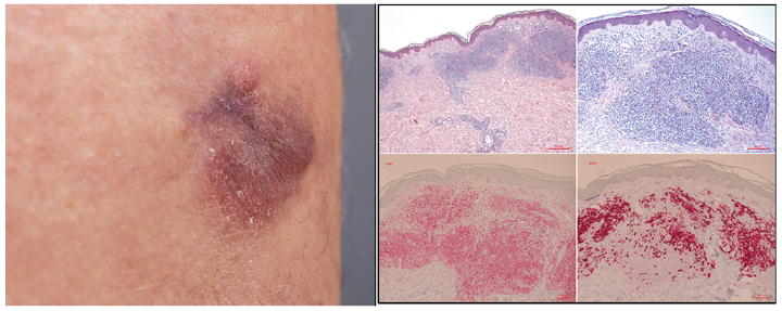
Fig. 1. Pseudolymphoma (a) Clinical picture of the skin. A 2 cm blue-reddish nodule on the right limb. (b) Histopathology. Dense infiltrate of lymphocytes in the upper dermis that are positive for T-(CD4) and B-(CD20) cell-markers (H&E × 50, × 100, CD4, CD20).
Fig. 2. Lymphadenosis cutis benigna. (a) Clinical features – 10 × 8 mm soft nodule at the helix of the right ear. (b) Histopathology: dense deep lymphocytic infiltrates affecting the deep dermis with CD4-positive Lymphocytes and CD20 positive B-Cells (H&E × 50, × 100, CD4, CD20).
Histopathology is an important tool in the diagnosis of CPL, and is necessary to distinguish CPL from reactive cutaneous inflammatory cell infiltrates, and lymphoma. No single histological feature alone can confirm the diagnosis of lymphoma or pseudolymphoma. Each sign is only more suggestive of a malign or benign behaviour of a neoplasm. Even the combination of histological features cannot confirm a CPL. Histopathologically CPL can be differentiated by the predominant cells in the infiltrate (Table I) (9, 10). In contrast to the WHO/European Organization for Research and Treatment of Cancer (WHO-EORTC) classification of cutaneous lymphoma, there is currently no established classification of pseudolymphomas.
Table I. Cutaneous pseudolymphoma can be differentiated according to the predominant cells in the infiltrate
Important histological characteristics of CPL that help to distinguish it from other benign cutaneous inflammatory cell infiltrates are the presence of variable numbers of medium- to large-sized lymphocytes, which may appear atypical, alongside many ordinary small lymphocytes and other inflammatory cells. Most CPLs consist of mixtures of B and T cells along with macrophages and dendritic cells (3).
Various histopathological features favour a lymphoma over a CPL:
However, CPL may be histopathologically indistinguishable from cutaneous lymphoma. This important differentiation may require additional tests, such as immunohistochemistry or gene rearrangement (3).
Immunohistochemistry differentiates lymphocytes by their immune-phenotype (surface markers), e.g. assignment to B or T cells. A mixed B- and T-cell infiltrate is more suggestive for CPL. A loss of typical T-cell-surface-markers (e.g. CD2, CD3, CD5), may be seen in progressed T-cell-lymphoma.
The diagnosis of CPL is backed by negative T-cell receptor rearrangement (monoclonality), missing plasma cell (mono-)clonality, negative immunoglobulin heavy chain gene rearrangements and polyclonal kappa and lambda light chains (3, 11–13).
Management of CPL may be difficult, with several treatment regimens, including topical and intralesional agents, systemic agents and physical modalities, showing variable dermatological responses.
We review here the treatment of pseudolymphoma of the skin according to its aetiology. We also suggest a treatment algorithm that may help physicians in choosing treatment. There is no previous systematic review of the treatment of CPL.
A systematic review was performed following the Preferred Reporting Items for Systematic Reviews and Meta-Analysis (PRISMA) based on PubMed, Medline and Web of Science databases using the query “skin pseudolymphoma treatment”. Our search was limited o “English” and “German” language, “human” subjects and publications from 1 January 1990 to 31 December 2015 documenting aetiology and/or adequate treatment for CPL. Furthermore, the references of the retrieved articles were searched manually for additional studies. The reference lists of the full-length articles were reviewed to identify additional articles meeting the pre-defined inclusion criteria. Articles in which pseudo-lymphoma was labelled as lymphocytoma cutis, lymphocytoma cutis benigna or lymphoid hyperplasia were included. Reviews and articles from the same department, in which some patients could have been described twice, were excluded. Table SI presents a summary of the characteristics of the included studies (author/study year, country, study design and number of patients). Data on sex/age, aetiology, time of onset, histological features, treatment and outcome were also collected. Descriptive statistics were used for the evaluation of sex/age and aetiology.
A total of 192 articles was identified from the initial search, of which 80 met the inclusion criteria. Three additional articles were found through the manual review. After searching in Medline and Web of Science databases, no further articles were added to the initial search. After reviewing all full-text articles and excluding reviews or papers about other diseases, a total of 83 studies; 80 from the initial search and 3 from additional papers, were analysed.
The results are based mostly on individual case reports and small case series. Treatment options should be decided first, according to CPL aetiology (Fig. 3). Treatment can be divided into topical and intralesional agents, systemic agents and physical modalities. Patient outcomes may vary, with the aetiology of the lesion playing an important role. Reported treatment regimens were often unsuccessful.
Fig. 3. Suggested classification and first- and second-line treatment for CPL. PUVA: psoralen and ultraviolet A therapy, UVA1: ultraviolet A1 therapy, ALA-PDT: photodynamic therapy with aminolevulinic acid, Top: topical treatment.
A total of 122 patients with pseudolymphoma of the skin were identified. The mean age of diagnosis was 42 years (n = 106), and the female:male ratio was 1:1 (n = 107). Data about the aetiology of the CPL were available for 106 patients (Fig. 4). The most frequently reported aetiology was CPL induced by drugs (n = 31), followed by tattoo reactions (n = 28).
Fig. 4. Aetiology of cutaneous pseudolymphoma (n = 106). SIT: specific immunotherapy.
Tattoo ink-related CPL. The literature review revealed 26 patients with CPL after tattooing. The pathogenesis remains obscure, but it is suggested that some metal components (cinnabar (red), cobalt salts (blue) and chromium salts (green)) are responsible for a delayed hypersensitivity reaction (8).
Surgery should be performed for tattoo ink-related CPL whenever possible. All the patients in our review who underwent a surgical procedure (n = 6) had complete healing, with no skin relapse or recurrence reported.
A total of 8 patients with CPL due to ink-tattoo reaction were treated with topical or intralesional corticosteroids. Six patients had a poor response after therapy, with one patient showing progression to frank lymphoma after 7 years (14–18). Only 2 patients experienced an improvement in the skin lesions (19, 20). One patient was successfully treated with topical corticosteroids in combination with oral hydroxychloroquine (21). Regular follow-up is recommended due to possible progression to malignancy.
Four patients with tattoo ink-related CPL were treated with laser. Q-switched neodymium:yttrium-aluminium-garnet laser or CO2 laser usually resulted in marked improvement in the skin lesions and in 2 of these 4 patients, intralesional corticosteroid adjuvant therapy was performed (17, 20, 22, 23).
Drug-associated CPL. Reports on 31 patients who developed drug-induced CPL were found. Medicaments responsible for causing CPL found in the review include: phenytoin, vancomycin, rifampicin, amlodipine, cefepime, sertraline, gabapentin, valsartan, etanercept, methylphenidate hydrochloride, lornoxicam, gemcitabine, carbamazepine, tricycle antidepressant, fluoxetine, H1 antagonist, H2 antagonist, phenothiazine and thioridazine. Magro et al. suggested that antihistamines may be associated with atypical lymphoid hyperplasia in some patients (24, 25). Other drugs that can be involved in cutaneous pseudolymphomatous reactions are angiotensin-converting enzyme (ACE) inhibitors, beta-blockers, calcium channel blockers, diuretics, cytotoxic agents (ciclosporin, methotrexate), rheumatology drugs (gold, salicylates, phenacetin, D-penicillamine, allopurinol, non-steroidal anti-inflammatory drugs (NSAIDs)), antibiotics, benzodiazepines, procainamide, oestrogens, progesterone and lovastatin (26). In a recent paper by Safa et al. (27), a female patient developed erythrodermic CD8+ pseudolymphoma during infliximab treatment and was treated successfully with ciclosporin after cessation of infliximab therapy. CPL may also appear in association with etanercept therapy (28).
In all patients with drug-associated CPL described here, the time of onset varied between 1 day and 15 years. The skin lesions resolved completely after drug discontinuation and the time period of regression ranged from a few weeks to few months.
Following therapy with medicinal leeches. Four patients developed CPL after therapy with medicinal leeches (Hirudo medicinalis). Medicinal leeches are used to treat chronic venous insufficiency, osteoarthritis and myalgia. Treatment with topical and/or intralesional corticosteroids resulted in improvement in the skin disease (29–32).
HIV-associated CPL. Two patients presented with a HIV-associated atypical cutaneous CD8+ T-cell infiltrate, which is usually associated with severe CD4 lymphopaenia (33). Its pathogenesis is unknown. One patient showed a decrease in the number of lesions after receiving systemic antiretroviral therapy with ritonavir, atazanavir, emtricitabine, tenofovir and oral prednisolone, and the other patient responded with a dramatic improvement in skin lesions after therapy with lamivudine, stavudine and indinavir (34, 35).
Borrelial lymphocytoma – lymphadenosis benigna cutis. This rare cutaneous manifestation of Lyme borrelioses appears in the early phase of infection, between 2 days and 6 months after a tick bite. Borrelial lymphocytomas are more common in children, and they are found mostly in the ear lobe, scrotum, and nipples (36). The gold standard treatment of borrelial lymphocytoma is treatment of the underlying infection with antibiotics, such as penicillin, doxycycline or cephalosporin. Only in rare cases, might other options apply: Aydogan et al. reported a male patient with borrelial lymphocytoma cutis resistant to penicillin and doxycycline treatment, who later responded to intralesional therapy with low-dose interferon-alpha-2a (37).
After immunotherapy, cat scratches, vaccination, injections, and arthropod bites. Yalcin et al. reported a case of Jessner’s lymphocytic infiltrate 3 weeks after initiation of bee venom immunotherapy, and 2 female patients developed B-cell pseudolymphoma shortly after initiation of a specific immune therapy (38, 39). In these cases, pseudolymphoma may resolve after immunotherapy is stopped.
Another case in a child suggested cutaneous lymphoid hyperplasia secondary to recurrent inoculation of Bartonella henselae due to feline scratches. The lesions responded to azithromycin and disappeared completely after the cat died (40).
Vaccine-induced CPL is rare, and its pathogenesis remains unknown, but some authors suggest that CPL may result from a delayed hypersensitivity reaction to vaccine components. This may be seen in vaccines containing aluminium, due to its contribution to a delayed absorption of other components, thereby promoting an antigen stimulus (41). In one patient, skin lesions disappeared after treatment with thalidomide 12 years after the onset of the disease in a patient with B-cell CPL after hepatitis B vaccination (42). In a case series of Cerroni et al., 4 female patients developed B-cell CPL after vaccination against early summer meningoencephalitis, tetanus and hepatitis B. Two patients were treated successfully with radiotherapy (43).
One patient with CPL associated with silicone injection and one with CPL at the site of a clonidine patch showed improvement of the lesions after treatment with triamcinolone acetonide injection.
Scabies mites may cause nodular scabies, histologically simulating mycosis fungoides or even cutaneous B-cell lymphoma. It has been postulated that this disease course occurs due to a delayed hypersensitivity reaction. Other arthropods may also cause this type of persistent reactions. Cases of skin pseudolymphoma caused by cutaneous leishmaniasis have also been reported (44, 45).
Lymphomatoid contact dermatitis. Having similar features with mycosis fungoides, but without Pautrier’s microabscesses, there are several allergens that may be implicated in lymphomatoid contact dermatitis. Treatment consists mainly in the avoidance of the causative allergens. Allergens implicated include phosphorus, N-Isopropyl-N-phenyl-4-phenylenediamine, ethylenediamine, dihydrochloride, cobalt naphthenate, nickel sulphate, paraphenylenediamine, gold sodium thiosulfate, zinc, squaric acid dibutylester, methylchloroisothiazolinone (26). Millican et al. reported a first case of CLH after sensitization with squaric acid dibutylester (46–48). Treatments include local and intralesional corticosteroids and tacrolimus ointments. Laser treatments or surgical excision may also be discussed with the patient. Successful surgical excision was performed in a case of CPL after intradermal testing with gold sodium thiomalate.
CPL in association with virus infections: herpes zoster, and molluscum contagiosum. Moreira et al. reported a patient with B-cell chronic lymphocytic leukaemia developing post-zoster CPL, which was treated with topical corticosteroids leaving a macular hyperpigmentation (49). Another case of CPL with this aetiology was published in 1987 (50). Surgical excision was performed in a female patient with CPL in association with molluscum contagiosum and the lesion did not recur (51).
Syphilis presenting as pseudolymphoma. With syphilis being the great imitator, clinicians should be aware of its many different clinical presentations and, especially, of a nodular form. A patient with nodular plaques with histological findings of a pseudolymphoma was treated with benzathine penicillin and all skin lesions cleared (52). Some cases of syphilis are clinically indistinguishable from lymphomatoid papulosis. Abundant plasma cells and spirochetes in the histology may help to differentiate them. Rash lesions resolve after treatment with antibiotics. Eleven other cases of syphilis have been reported to histologically mimic a malignant lymphoid neoplasm (52, 53).
Actinic reticuloid. This chronic photodermatitis, usually affecting male older patients, is characterized by extreme photosensitivity. It is suggested that, due to alterations induced by a photoallergic reaction, normal components of the skin change into “antigens”. There is no cure, and sun-protection is recommended (54, 55).
Psoralen plus ultraviolet A and long-wave ultraviolet A therapy. A recent report showed good results in a patient with lymphocytoma cutis (Spiegler-Fendt sarcoid) after therapy with 13 sessions of psoralen plus ultraviolet A (PUVA) and 5 sessions of intralesional triamcinolone (56). In another case, PUVA was the therapy of choice of a solitary T-cell pseudolymphoma of the breast (57). Long-wave ultraviolet A (UVA1) in a young man having palpable migratory arciform erythema led to complete regression of the skin lesions (58).
5-aminolevulinic acid photodynamic therapy. Treatment with ALA-PDT was reported in 4 patients with CPL of the face, leading to marked improvement in the skin lesions. In 1 patient there was no recurrence (59–61).
Topical tacrolimus. Two patients with lymphocytoma cutis of the head and neck with idiopathic aetiology showed slow clinical improvement (>4 months) of the affected skin while using topical tacrolimus 0.1% (62). Nevertheless, spontaneous regression of CPL was reported (63).
Topical imiquimod. This immunomodulator (Toll-like receptor 7 inhibitor) may be an efficient treatment alternative for CPL. Topical treatment with imiquimod 5% resulted in total clearance of the affected skin after treatment failure with topical steroids, antimalarials combined with tacrolimus 0.1%, photodynamic therapy, ultraviolet B (UVB) and oral penicillin (64).
Hydroxychloroquine sulphate. Treatment with hydroxychloroquine sulphate (400 mg/day) for 3 months was successful in an individual with pseudolymphoma cutis without any causal factor, and this patient remained disease-free at one year of follow-up (65). One woman, having miliarial-type perifollicular B-cell pseudolymphoma, had an improvement of the skin lesions after treatment with hydroxychloroquine (66). A hypothesis providing a rationale for treatment with hydroxychloroquine is the presence of plasmacytoid dendritic cells in CPL, as these cells respond to hydroxychloroquine (67). We suggest the use of hydroxychloroquine in patients with CPL with unknown aetiology in whom excision cannot be performed. However, dermatologists should be aware that this recommendation is based on only a few case reports.
CPLs are a group of diseases that exhibit a lymphocyte-rich infiltrate, which either clinically and/or histologically simulate cutaneous lymphomas (68).
A wide range of causative agents has been described, and treatment may thus be difficult. Many different modalities have been reported. Following a systematic review of the literature of the last 25 years we suggest that treatment options should be chosen based on the specific aetiology (Fig. 4). While there are a considerable number of publications on the histopathology and classification of CPL, reports on treatment options are lacking, with much of the published work not disclosing any information about successful treatments and (long-term) outcomes. More reports of cases of CPL are needed, whether successfully treated or not, in order to avoid publication bias under-reporting poor outcomes or overestimating the response rates. Randomized controlled trials are difficult due to the rarity of CPL, and a gold standard therapy cannot be suggested. Treatment choice should be made individually for each patient according to aetiology and localization. A large proportion of cases of CPL are idiopathic and, if no surgical excision is performed, watchful follow-up is highly recommended due to the possible progress of CPL to malignant lymphoma.
The authors have no conflict of interest to declare.


 Click to show fullsize
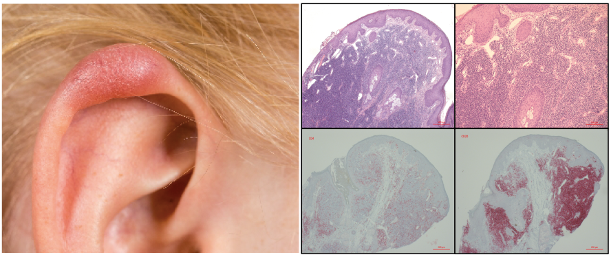 Click to show fullsize
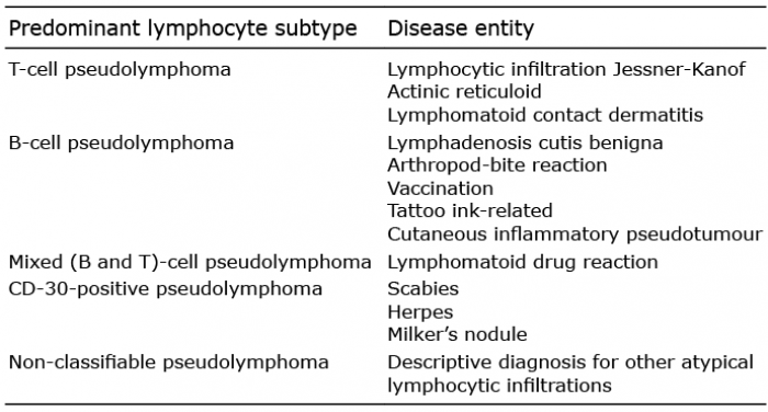 Click to show fullsize
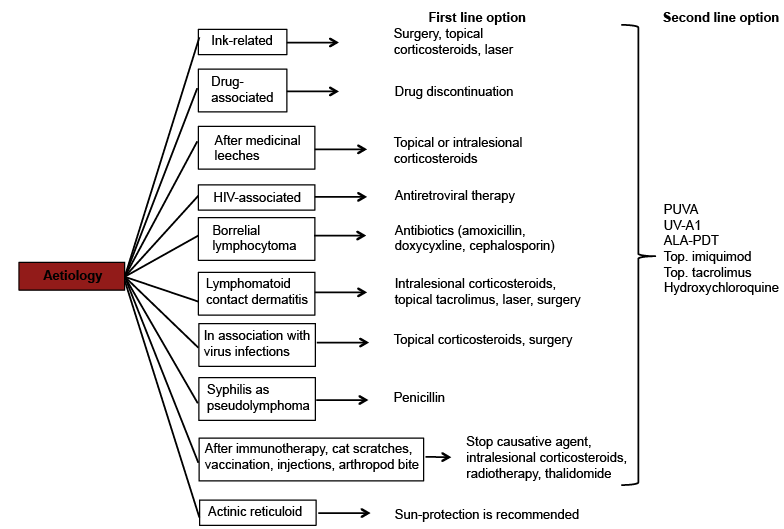 Click to show fullsize
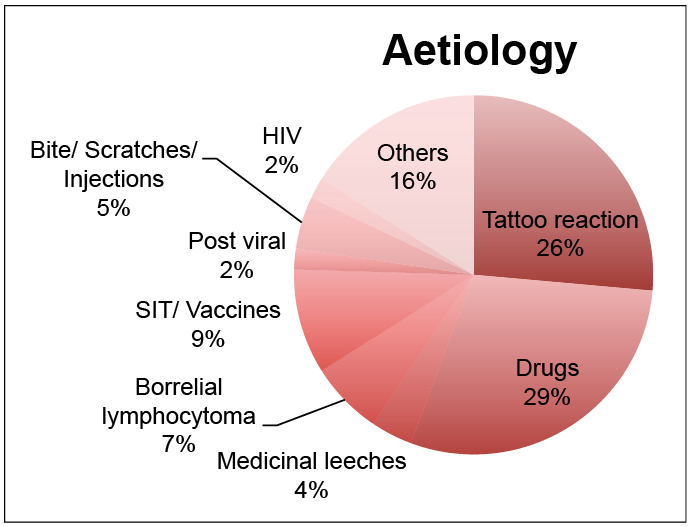 Click to show fullsize