


1Department of Dermatology and Venereology and 5Department of Obstetrics and Gynaecology, Lund University, Skåne University Hospital, Malmö, 2Regional Cancer Centre Uppsala-Örebro, Uppsala University Hospital, Uppsala, 3Department of Medical Microbiology Laboratory Medicine, Lund University, Lund, and 4Department of Urology, Helsingborg General Hospital, Lund University, Helsingborg, Sweden
Studies on risk factors for penile intraepithelial neoplasia have been small in size, have not distinguished penile intraepithelial neoplasia from invasive cancer, and have relied on self-reported information. This study investigated risk factors for penile intraepithelial neoplasia in a cohort of 580 penile intraepithelial neoplasia cases and 3,436 controls using information from 7 Swedish registers. Cases with penile intraepi-thelial neoplasia had increased odds ratios (ORs) for inflammatory skin diseases (14.7, 95% CI 6.5–33.4) including lichen planus (12.0, 95% CI 3.0–48.0), indicating lichen planus to be an important risk factor. Increased ORs were also observed for diseases of the prepuce (4.0, 95% CI 2.2–7.4), immunosuppressive drugs (5.0, 95% CI 2.5–9.8), penile surgical procedures (4.8, 95% CI 2.2–10.8), balanitis (9.2, 95% CI 5.0–16.8), genital warts (9.9, 95% CI 4.3−22.7) and organ transplantation (7.0, 95% CI 2.4–20.8). This study demonstrates important risk factors for penile intraepithelial neoplasia, providing knowledge that can help prevent the development of penile cancer.
Key words: PeIN; penile intraepithelial neoplasia; penile cancer in situ; CIS; penile cancer; risk factors: lichen planus.
Accepted Nov 14, 2018; E-published Nov 14, 2018
Acta Derm Venereol 2019; 99: XX–XX.
Corr: Sinja Kristiansen, Department of Dermatology and Venereology, Skåne University Hospital, SE-214 28 Malmö, Sweden. E-mail: Sinja.Kristiansen@med.lu.se
Non-invasive penile cancer is a precursor to invasive penile cancer. Studies on risk factors have been small in size and have relied on self-reported information. This study looked at risk factors for non-invasive penile cancer in 580 men and 3,436 cancer-free controls in Swedish medical registers. It found that inflammatory skin diseases, diseases of the prepuce, taking immunosuppressive drugs, having had penile surgery, inflammation of the glans, genital warts and being organ transplanted all are risk factors for non-invasive penile cancer. Increased knowledge on risk factors for non-invasive penile cancer will help in the prevention of development of invasive penile cancer.
Penile cancer is a rare type of cancer with an age-adjusted incidence in Sweden of 2.1/100,000 (1). In Europe, the incidence is 0.45–1.7/100,000 (2), and in the USA 0.58/100,000 (3). The highest incidence is reported from Brazil, with 2.9–6.8/100,000 (4), and Uganda with 3.3/100,000, although in Uganda recent data has shown a significant decrease in incidence to 1.2/100,000 (5). In contrast, increasing incidence is seen both in the Netherlands (6) and the UK (7). In Sweden, the incidence was stable between 2000 and 2012 (1). In more than 95% of cases, penile cancer is classified as squamous cell carcinoma (SCC) (8). Penile intraepithelial neoplasia (PeIN) is a premalignant precursor lesion of invasive penile SCC, a SCC in situ, where the squamous epithelium shows dysplastic changes with an intact basement membrane (9). Each year approximately 150 penile cancer cases are diagnosed in Sweden, approximately 30% are diagnosed with PeIN (1). PeIN are morphologically divided into 4 subgroups, with differentiated PeIN being the most predominant, followed by warty-basaloid, basaloid and warty morphology (10). In 2016, the World Health Organization (WHO) introduced a new pathological classification of PeIN based on aetiology, with 2 main pathways for malignant transformation, one pathway being a human papillomavirus (HPV)-induced carcinogenesis, named “undifferentiated PeIN”, and the other being an inflammatory pathway derived from lichen sclerosus (LS) and lichen planus (LP), named “differentiated PeIN” (9, 11). Data on how often premalignant lesions transform to invasive cancer are scarce. According to published data the transformation occurs in 10–30% of cases in PeIN, with the glans and inner prepuce having the highest risk of malignant transformation (12, 13). The overall 5-year relative survival rate in Sweden is 82% (1).
The prevalence of HPV in manifest penile cancer is approximately 47%, ranging between 24 and 82% (14, 15). HPV is assumed to be a major risk factor for penile cancer. In PeIN, HPV prevalence is more common, between 87–100% of cases (16, 17). Inflammatory skin disease, such as LS, is present in 28–55% of cases of penile cancer and is thought to be the other major risk factor for invasive penile cancer (18–20). Balanitis and phimosis increases the relative risk of invasive penile cancer 9.5 times (21). Circumcision in infancy is known to protect from developing penile cancer (22, 23). Smoking is associated with a 4.5% increased risk of invasive penile cancer (22). Overweight and obesity are more prevalent among patients with invasive penile cancer, indicating these to be risk factors (24). Psoralen plus ultraviolet A (PUVA) treatment has been shown to be a risk factor for penile cancer, with an incidence rate ratio of 4.5 (95% CI 1.3–16.1) (25). Immunosuppression following organ transplantation increases the risk of non-melanoma skin cancer (NMSC), PeIN and invasive penile cancer (26, 27). Socioeconomic factors, such as low educational level, low disposable income and being single, are also known risk factors for invasive penile cancer, but not for PeIN (28, 29).
Risk factors for PeIN, as distinct from invasive penile cancer, have not been well studied. Previous studies are small in size, controls are seldom used, and data on risk factors are often based on self-reports. Knowledge of risk factors for PeIN is important to identify preventable causes for PeIN.
The aim of the present study was to investigate risk factors for PeIN, using information registered by medical doctors, in a large cohort of 580 Swedish men with PeIN and 3,436 controls, between the years 2000 and 2012.
The Swedish National Penile Cancer Register (NEPCR) was founded in the year 2000 and all penile cancers are registered, with a coverage of > 99% of those registered in the mandatory Swedish Cancer Register (SCR). In 2013, a database, Penile Cancer Data Base Sweden (PenCBaSe), was generated by the Steering committee of the NEPCR in collaboration with the Swedish Regional Cancer Centre, linking the NEPCR with the National Inpatient Register (IPR), the National Outpatient Register (OPR), the SCR, the Cause of Death Register, the National Register of Prescribed Drugs (RPD), the Register of the Total Population and the Longitudinal Integration Database for Health Insurance and Labour Market Studies, respectively. The IPR provides information on inpatient care and was founded in 1964 with coverage of the whole nation since 1987. In 1997, the OPR was founded and covers outpatient surgeries, and since 2001 the OPR has also included all hospital-based outpatient physician visits (30). The RPD was founded in 2005 and holds information on all prescribed drugs that patients obtain from pharmacies. The registers are further described in a study published previously (28).
The current International Classification of Diseases (ICD) in Sweden is the ICD-10 (for the complete list of all ICD codes searched, see Table SI), established in 1997. The Anatomic Therapeutic Chemical (ATC) classification system was searched for medication with immunosuppressive drugs (for a list of codes see Table SI).
In the database PenCBaSe, generated in 2013, all diagnoses of PeIN in NEPCR between 2000 and 2012 were included. Six controls without penile cancer diagnosis were randomly chosen for every case of PeIN, matched on year of birth and county of residence. All cases and controls in the PenCBaSe database were included in the current study. Educational level was divided into 3 different groups: “low”, meaning up to 9 years of school, “middle”, meaning 10–12 years of school, and “high”, meaning more than 13 years of school. Comorbidity was estimated using the validated Charlson Comorbidity Index (CCI) (31). Disposable income as a measure of socioeconomic position was divided into 2 groups, above and below the median for controls, calculated to be representative for the population. For each case and all controls, registers were searched for diagnoses, surgical procedures and immunosuppressive medication, in each case checking back to the start of each register.
Registers were searched for diseases of the prepuce, such as phimosis and paraphimosis, medication with immunosuppressive drugs, balanitis, genital warts, organ transplantation, inflammatory skin diseases, such as LS and LP, surgical interventions on the penis, smoking, tobacco use, chronic obstructive pulmonary disease (COPD) and obesity. Smoking and use of tobacco were seldom registered; hence, the incidence of smoking was estimated through looking at the diagnosis of COPD. All diagnoses, surgical procedures and immunosuppressive drugs given within one year prior to the PeIN diagnosis were excluded to ensure the diagnoses were not misclassified PeIN diagnoses. Registers were also searched for body mass index (BMI), psoralen plus ultraviolet A (PUVA) treatment, photodynamic therapy (PDT), genital itch, HIV and HPV-related cancers such as head, neck and anal cancer, but due to the low number, these diagnoses were excluded. Circumcision is seldom performed in infancy in Sweden. Circumcision in adult men is performed mostly because of phimosis.
The study was approved by the Swedish ethical board in Lund (Dnr 2015/907).
Conditional logistic regression was used to compare the risk of PeIN selected risk factors. Crude odds ratios, as well as odds ratios adjusted for educational level, marital status and comorbidity, were calculated. Complete case analysis was used, since the data were extensive with a small proportion of missing information.
A sensitivity analysis was performed excluding diagnoses at 0, 6 and 12 months, respectively (see Table SII). All statistical analyses were performed using the R program package (3.4.3) by R Core Team (2017).
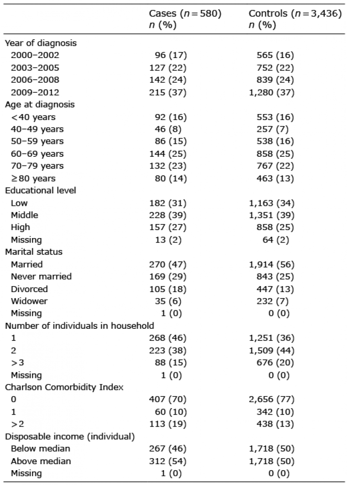
A total of 580 male patients with PeIN and 3,436 male controls matched on age and county were included in the study. Median age was 64 years (range 17–94 years) and 64 years (range 16–95 years) for the cases and controls, respectively (Table I). The vast majority of the cases and controls were Caucasian, with only 3.8% (22/580) of cases and 4.7% (161/3436) of controls born outside Europe (data not shown).
Being married was more common among controls than among cases (56% vs. 47%). The cases were more often never married or were divorced. Among the cases, 46% were living in a 1-person household compared with 36% of controls. Nineteen percent of cases had a CCI of 2+ compared with 13% of controls. In disposable income above the median, no difference was observed between cases and controls (54% vs. 50%). Of the controls, 34% had a low level of education compared with 31% of the cases, showing no difference in the level of education.
Table I. Cohort characteristics for cases and controls
Table II shows all diagnoses, penile surgical procedures and immunosuppressive drugs registered for the cases and controls up to one year prior to the cases’ PeIN diagnoses. Diseases of the prepuce, meaning phimosis, paraphimosis and adherent prepuce, were found in 3.1% of cases and in only 0.8% of controls (p < 0.001). Immunosuppressive drugs were taken by 2.8% of cases compared with 0.6% of controls (p < 0.001). Among the cases, 1.9% had gone through a penile surgical procedure before the PeIN diagnosis, compared with only 0.4% of controls (p < 0.001). Balanitis was found in 4.7% of cases compared with 0.5% of controls (p < 0.001). Genital warts were found in 2.6% of cases, but in only 0.3% of controls (p < 0.001). Organ transplantation was found in 1.2% of cases compared with 0.2% of controls (p < 0.001). LS and LP were found in 3.4% of cases compared with 0.2% of controls (p < 0.001).
Obesity was observed in 1.6% of cases compared with 0.7% of controls (p = 0.03). Smoking and tobacco use were registered in 0.5% of cases and 0.3% of controls, showing no statistical difference (p = 0.44). COPD was diagnosed in 1.4% of cases and in 1.5% of controls, with no statistical difference (p = 0.79).
Table II. Diagnoses, procedures and drugs found until 1 year prior to cases’ penile intraepithelial neoplasia diagnosis
Fig. 1 shows the crude odds ratios (ORs) one year prior to the PeIN diagnoses. Men with the following diagnoses had an increased OR for PeIN compared with the controls; diseases of the prepuce OR 4.0 (95% confidence interval (CI) 2.2–7.4, p < 0.001), patients medicating with immunosuppressive drugs OR 5.0 (95% CI 2.5–9.8, p < 0.001), surgical procedures on the penis with an OR of 4.8 (95% CI 2.2–10.8, p < 0.001), balanitis OR 9.2 (95% CI 5.0–16.8, p < 0.001), genital warts, OR 9.9 (95% CI 4.3−22.7, p < 0.001), organ transplantation OR 7.0 (95% CI 2.4–20.8, p < 0.001), LS OR 13.6 (95% CI 5.2–35.5, p < 0.001) and LP OR 12.0 (95% CI 3.0–48.0, p < 0.001).
In obesity an OR of 2.4 (95% CI 1.1–5.3, p = 0.03) was calculated. No statistically significant difference between cases and controls was shown for either smoking/tobacco use (OR 1.7, 95% CI 0.5–6.1, p = 0.44) or COPD (OR 0.9, 95% CI 0.4–1.9, p = 0.79).
Fig. 1. Forest plot with odds ratio (OR) and 95% confidence interval (95% CI) for diagnoses, procedures and drugs, one year prior to the cases receiving a diagnosis of penile intraepithelial neoplasia.
The ORs were adjusted for marital status, educational level and CCI (Table III). Further adjustment for the number of individuals in the household, disposable income, and country of birth did not significantly alter the ORs (data not shown).
Table III. Crude and adjusted odds ratios (OR) for marital status, Charlson Comorbidity Index and educational level
This extensive Swedish case-control study found a strong OR in men diagnosed with PeIN for LP (12.0) which has never been shown previously. In addition, this study observed increased ORs in alignment with previous studies for diseases of the prepuce (OR 4.0), immunosuppressed patients (OR 5.0), prior surgical procedures on the penis (OR 4.8), balanitis (OR 9.2), genital warts (OR 9.9), organ transplantation (OR 7.0) and LS (OR 13.6).
Furthermore, the current study showed that never having been married or being divorced were risk factors for PeIN, but no difference between the cases and controls was observed for low disposable income and low education. This is consistent with Torbrand et al. (28) who did not observe any association between PeIN and low educational level or low disposable income. A Danish population-based study of men with penile cancer found an increased risk of invasive penile cancer in single men compared with married men, but no data on PeIN was shown (29).
In the current study, LS was diagnosed in 2.4% of PeIN cases with an OR of 13.6 at least one year prior to the PeIN diagnosis. To our knowledge no study has investigated preceding LS in PeIN patients. Pietrzak et al. (18) found LS in 29% (10/34) of pathology reports of consecutive PeIN cases investigated at one centre, but our finding of an increased OR of 13.6 for LS diagnosed one year prior to the PeIN diagnosis, indicates LS as an important risk factor for PeIN. In invasive penile cancer, at the time of diagnosis of the invasive cancer, LS was found in 33% (68/207) in a study 2003 by Velaquez & Cubilla (20), and Powell et al. (19) reported the highest frequency of 55% (11/20) of LS in 20 penile pathology SCC reports.
The malignant risk of penile LP among those that have developed penile cancer has not been well studied. The main research comprises case reports, although Mannweiler et al. (11) found 9 cases of penile cancer with concomitant LP in pathology reports of 35 HPV negative cancers. In the current study, LP was more common in PeIN (1.0%, OR of 12.0), than in controls, indicating that LP might be an important risk factor for PeIN.
Balanitis was seen more often in PeIN cases (4.7%, OR of 9.2) compared with controls in the current study. No previous study has demonstrated any association with balanitis and PeIN. We found one study by Maden et al. (32) in which no association between balanitis and PeIN was reported. In invasive penile cancer Hellberg et al. (21) showed an increased relative risk of 9.5 (95% CI 5.2–17.2) for balanitis, which declined to 5.22 after adjustment for phimosis, corroborating our results of an OR of 9.2 for PeIN.
HPV status was not registered in NPECR, limiting the information about HPV both in the cases and controls. Instead, diagnosis of genital warts was used as a predictor of HPV, but this most likely rendered an underestimation of HPV, since only approximately 19% of males with genital low-risk HPV types develop genital warts (33). Genital warts were diagnosed in only 2.6% of our cases (OR of 9.9), but in several studies high-risk HPV types were detected in up to 100% of PeIN at the time of diagnosis (16, 17).
Circumcision in infancy is known to be highly protective against the development of penile cancer. However, the effect on PeIN is not as strong (22, 23). Israel has the lowest incidence of penile cancer in the world, with only 0.1 per 100,000, due to a high rate of circumcision in infancy (2). In contrast, circumcision performed later in life has been shown to be associated with an elevated risk of penile cancer (34). For Swedish men most excisions on the penis are performed due to problems with retracting the foreskin, in most cases expected to be caused by phimosis, LS or LP. Our findings of an increased OR for penile surgical procedures suggests a risk of PeIN that is probably due to phimosis, LS or LP.
In the current study, 1.2% of the PeIN cases were organ-transplanted (OR 7.0) and 2.8% obtained immunosuppressive drugs (OR 5.0). Organ-transplanted patients have to be on lifelong immunosuppressive medication. Immunosuppression is a known risk factor for NMSC (26). Our finding is consistent with Madeleine et al. (27), who showed that organ-transplanted patients had an increased standardized incidence ratio (SIR) for PeIN of 18.6 and an SIR for invasive penile cancer of 3.9.
In the current study, obesity was seen in 1.6% of PeIN cases (OR 2.4). Literature on obesity and PeIN could not be found and data on obesity and invasive penile cancer is scarce. Barnes et al. (24) demonstrated that invasive penile cancer cases were significantly more overweight (OR 2.64) and obese (OR 3.24) compared with controls. Our results, with an OR of 2.4 (CI 1.1–5.3, p = 0.03) for obesity, indicate that obesity can be a potential risk factor for PeIN, but this needs to be studied further.
A major strength of this study is the size, since it includes as many as 580 PeIN cases and 3,436 matched controls. Another strength is that the diagnoses of potential risk factors used for analysis were made and registered by medical doctors at least 1 year before the PeIN diagnoses. An additional strength is that Sweden has nationwide medical registers with excellent coverage, making it possible to study several parameters and diseases that are potential risk factors for PeIN over time.
A limitation of the study is that some of the interpreted risk factors could have been misclassified PeIN diagnoses, since diagnoses for risk factors were not always confirmed by a pathological evaluation. To minimize the risk of misclassification, a sensitivity analysis was performed excluding different lapses of time for diagnoses of potential risk factors, and the statistically significant differences persisted regardless of the length of these lapses (see Table SII).
Smoking has been shown to be associated with a 4.5% increased risk of invasive penile cancer (22). Furthermore, a dose relation has been reported, with a relative risk of having penile cancer of 1.88 (95% CI 1.10–3.19) for men smoking more than 10 cigarettes/day (21). A limitation of the current study is that smoking is not regularly coded in Swedish registers, leading to very few registered codes, with no statistical difference between cases and controls. Instead, the code for COPD was used as a proxy for smoking, but since it takes many years to develop COPD, this is probably an underestimation of smoking as a risk factor for PeIN, and no differences were shown for COPD between the cases and controls in the current study.
This study showed statistically significant increased OR in PeIN patients for several known risk factors that have not been well described previously, such as diseases of the prepuce, balanitis, genital warts, LS and surgical procedures on the penis. It also found a significant increased OR of 12.0 in PeIN patients for LP, suggesting LP to be an important risk factor for PeIN. To our knowledge this has not been reported previously. This study has identified preventable risk factors for PeIN. This knowledge can be used to prevent invasive penile cancer. Important steps for governments and medical doctors to implement towards prevention of PeIN are HPV vaccination programmes for prepubescent boys, vaccination before organ transplantation, and treatment and follow-up of men with penile inflammatory skin diseases.
The authors have no conflicts of interest to declare.


 Click to show fullsize
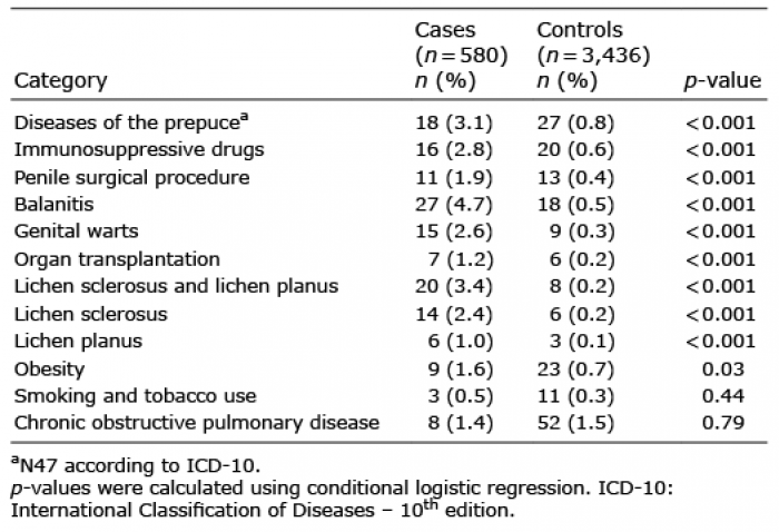 Click to show fullsize
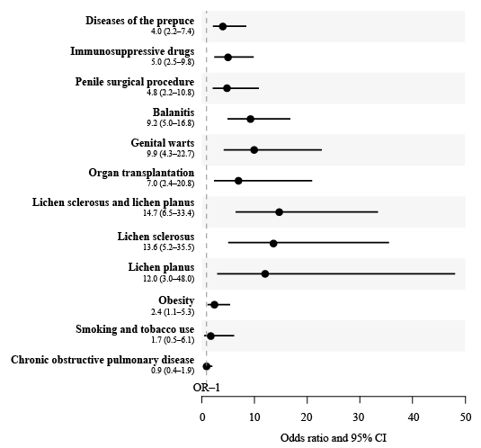 Click to show fullsize
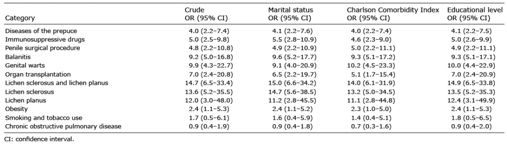 Click to show fullsize