


1Department of Dermatology, Faculty of Medicine and Graduate School of Medicine, Hokkaido University, N15 W7, Kita-ku, Sapporo 060-8638, and 2Department of Bacteriology, Hokkaido Institute of Public Health, Sapporo, Japan. *E-mail: yfujita@med.hokudai.ac.jp
Accepted Jan 16, 2019; E-published Jan 17, 2019
Septic vasculitis or septic vasculopathy is defined as vascular changes occurring in patients with sepsis (1). Although the process whereby the cutaneous changes develop has yet to be fully clarified, several complex pathogenic mechanisms of septic vasculitis are assumed, such as disseminated intravascular coagulation, direct vessel wall invasion by the microorganism, immune-mediated vasculitis and septic embolism (2). Although these have been observed histopathologically, there have been no investigations of whether the toxins directly affect skin vessels (1). We present here a case of septic vasculitis that preceded thoracic empyema, in which deposition of Staphylococcus aureus enterotoxin (SE) on the vessel walls was a factor in the severe cutaneous manifestations. This case suggests that SE plays a significant role in the pathogenesis of septic vasculitis.
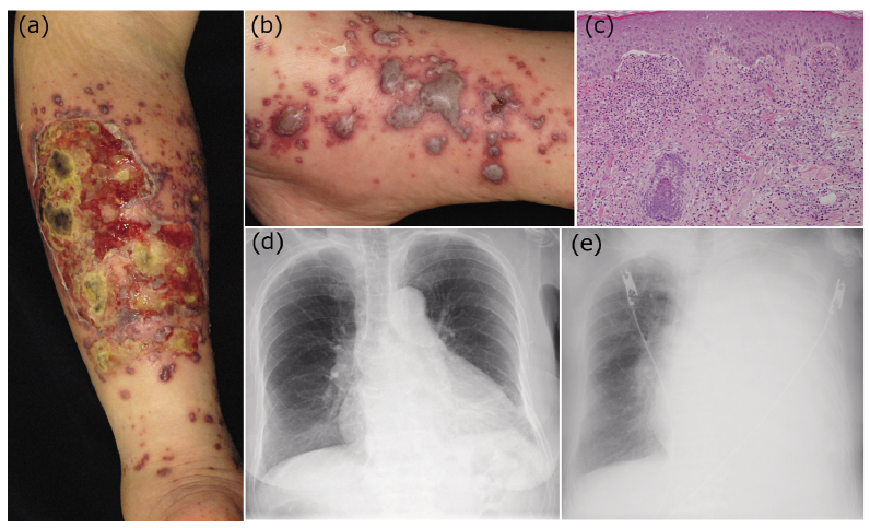
An 82-year-old Japanese woman was referred to our department with a 1-week history of necrolytic ulcers on the lower legs. Physical examination revealed reddish-violaceous ulcers, 15 cm in size, with yellow or black necrolytic tissue surrounded by disseminated blood blisters and purpura on the flexor surfaces of the lower legs (Fig. 1a, b). A biopsy specimen obtained from purpura showed interface changes, dermal neutrophilic infiltrates, extravasated erythrocytes and small blood vessel thrombi (Fig. 1c). Ziehl-Neelsen, Periodic acid–Schiff, Grocott and Gram-staining were all negative. The patient’s vital signs at administration (day 0) were as follows: body temperature 38.1°C, heart rate 115 beats per min, blood pressure 129/76 mmHg, respiratory rate 16 breaths per min and oxygen saturation as detected by the pulse oximeter (SpO2) 93% in room air. The patient’s subjective symptoms were unremarkable. She had a medical history of dementia. Laboratory examinations revealed a white blood cell count of 13,800 mm3 (88% neutrophils) and C-reactive protein of 20.43 mg/dl (normal range 0–0.39). All antibodies were negative, including anti-glomerular basement membrane antibodies, anti-neutrophil cytoplasmic antibodies, anti-desmoglein 1/3 antibodies and anti-BP180 antibodies. A chest X-ray showed mild infiltrates in the left lower lung (Fig. 1d). A transthoracic echocardiogram showed no vegetation. Intravenous ceftriaxone (1 g every 24 h) was administered after blood culture. On day 2, methicillin-susceptible S. aureus (MSSA) was identified from blood samples, and the antibiotics were switched to ampicillin/sulbactam (3 g every 12 h). However, her respiratory condition worsened in 3 days. Chest X-ray on day 5 showed massive pleural effusion in the left lower lung (Fig. 1e), which led to chest tube insertion to treat thoracic empyema. Since the culture from the pleural effusion also yielded MSSA, the time interval of antibiotics administration was shortened to every 6 h. Thereafter, the drainage volume decreased and ulcers on the lower legs gradually epithelized, and the patient was discharged on day 30. From this disease course, the diagnosis of septic vasculitis that preceded thoracic empyema was made.
Fig. 1. (a) Erythematous-violaceous ulcers, 15 cm in size, with yellow or black necrolytic tissue surrounded by disseminated multiple blood blisters and purpura on the flexor surfaces of both lower legs. (b) Many blood blisters of various size on the right ankle. (c) A biopsy specimen obtained from purpura shows interface changes, dermal neutrophilic infiltrates, extravasated erythrocytes and small blood vessel thrombi (haematoxylin and eosin staining, original magnification ×40). (d) A chest X-ray at admission shows infiltrates in the left lower lung. (e) A chest X-ray shows massive pleural effusion in the left lower lung 5 days after admission.
To investigate the pathogenesis of the cutaneous lesions, we analysed the biological properties of MSSA colonies obtained from 2 independent blood cultures and 2 independent pleural effusion cultures. PCR detecting genes encoding SE serotypes A-R, performed according to the method of Omoe et al. (3), identified staphylococcal enterotoxin-like toxin type P (SElP) in all specimens (Fig. 2a). Sandwich enzyme-linked immunosorbent assays from SE strains confirmed the production of SElP (16.1 ng/ml, 16.6 ng/ml from 2 independent blood cultures and 18.1 ng/ml, 18.3 ng/ml from 2 independent pleural effusion cultures, negative control <0.25 ng/ml) (4). Reverse passive latex agglutination reaction assays specific for toxic shock syndrome toxin 1 (TSST-1), exfoliative toxin and Panton-Valentine leucocidin (PVL) were negative (5–7) (data not shown).
Since the histopathology showed no microorganisms, it was suspected that MSSA toxins directly affected the vessels in the present case. Therefore, direct immunofluorescence of the lesional skin to visualize the deposition of SE was performed (Appendix S1). Deposition of SE within the endothelial cells was identified in a cutaneous specimen from a purpura (Fig. 2b), whereas no SE deposition was observed in the skin of a healthy control, in the lesional skin of cutaneous small vessel vasculitis, or in the non-lesional area of the current patient (data not shown). Furthermore, this negativity was confirmed in these sections without the primary antibody. These results indicate that SE deposition on the vessel epithelium was causative of septic vasculitis in the present case.
Fig. 2. (a) Detection of the SElP gene by multiplex PCR (4). Lanes: M, molecular size marker; B1 and B2, colonies from blood culture; P1 and P2, colonies from pleural effusion culture; P, a mixture of total DNA of S. aureus 196E, S6, FRI-326, FRI-569 and N315 as a positive control; N, distilled water as a negative control. (b) S. aureus enterotoxin (SE) deposition (arrowheads) within pericytes of affected vessels in a cutaneous specimen from a purpura (green: SE; blue: nuclei, original magnification ×200).
Septic vasculitis in the skin manifests as purpuric papules, plaques, petechiae, vesicles, bullae and pustules, which generally appear in the early stage of systemic sepsis (1, 8, 9). The current case presented widespread cutaneous manifestations that preceded the diagnosis of thoracic empyema symptoms by 2 weeks. Without hospitalization based on the cutaneous findings, the diagnosis of empyema might have been delayed due to the patient’s poor subjective symptoms due to dementia. The prodromal cutaneous lesions resulted in the rapid and appropriate systemic interventions of the present case.
Different microorganisms can cause septic vasculitis (1, 8, 9). In our case, MSSA was the causative agent. S. aureus has many superantigens, such as TSST-1, TSST-1-like toxin, exfoliative toxin, PVL and various SEs (10, 11). SE is known to be associated with purpura fulminans in the dermatological field; however, there have been few investigations on the serotype of SEs (12). To date, there have been no investigations on the effect of SElP on the skin. In the present case, the causative MSSA produced SElP exclusively, which indicated that SElP played a significant role in the pathogenesis of the skin lesions. SElP is known to have an emetic activity and to act as a superantigen (4). SElP induces the secretion of interleukin-2, 4, interferon-gamma, and tumour necrosis factor-alpha from human T cells (4). which might cause systemic cytokine storm resulting in immune-mediated vasculitis and intravascular coagulation in the skin.
In addition, the radical progression of purpura preceding empyema suggests that there was systemic circulation of SE, and that this circulation directly affected vasculitis formation. Indeed, we first identified deposition of SE within pericytes of affected vessels by direct immunofluorescence. The direct deposition of SEs may explain the trigger of angiotropic infiltration of inflammatory cells resulting in cutaneous vasculitis.
Dermatologists should take note of cutaneous vasculitis followed by severe systemic infection with a gap in the clinical course. Although the precise mechanism of septic vasculitis remains to be elucidated, this case report provides a clue to the pathogenesis from the viewpoint of SE.
The authors thank Dr Nozomu Takei, Dr Ai Sawaoka, Dr Kentaro Nagaoka, Dr Masaru Suzuki and Professor Satoshi Konno (Department of Respiratory Medicine, Faculty of Medicine and Graduate School of Medicine, Hokkaido University, Japan) for the treatments of empyema, and Professor Katsuhiko Omoe (deceased, Department of Veterinary Medicine, Faculty of Agriculture, Iwate University, Japan) for the experimental methods of SE.
The authors have no conflicts of interest to declare.


 Click to show fullsize
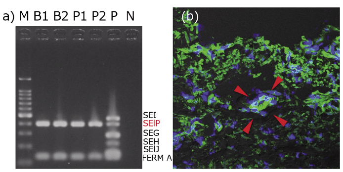 Click to show fullsize