


Department of Dermatology, Peking Union Medical College Hospital, Chinese Academy of Medical Sciences and Peking Union Medical College, Dongcheng District, Beijing 100730, China. *E-mail: zengyueping0917@126.com
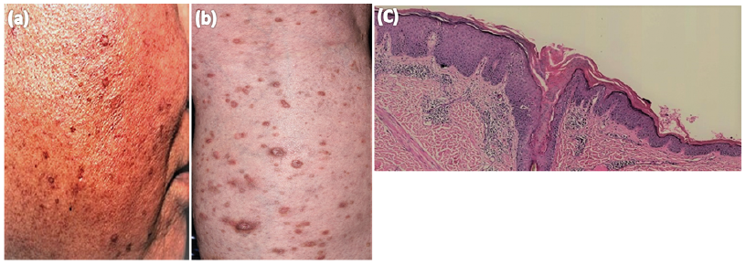
Case 1. A 64-year-old man presented with scattered cyclic rashes with a slightly raised edge on his face, which had developed 4 years earlier (Fig. 1a). In addition to pre-existing lesions, generalized discrete, sharply demarcated, brownish-red coloured, somewhere excoriated, macules and flat papules, 3–10 mm in diameter had developed on his trunk and extremities one year previously (Fig. 1b). The pruritic papules did not respond well to topical steroid ointments.
Case 2. A 68-year-old man presented with multiple bean-sized red flat papules with mild itching on both his lower extremities (Fig. 2a). The itching relieved on treatment with topical halometasone, but the skin lesions did not resolve. He reported that he had recurrent annular macules with no obvious scaling on his palms for the past 10 years (Fig. 2b), which were relieved on treatment with topical halometasone ointment.
In both cases, there was no family history of similar diseases. Routine serological tests were unremarkable. Skin biopsies were taken from reddish papules on the patients’ thighs.
What is your diagnosis? See next page for answer.
Fig. 1. (a) Scattered cyclic rashes with a slightly raised edge on the face. (b) Generalized discrete, sharply demarcated, brownish-red coloured macules and flat papules on the leg. (c) Histological examination revealed hyperkeratosis, multiple columns of parakeratosis overlaying an epidermal depression (cornoid lamella), with a thin or absent granular layer and scattered dyskeratotic keratinocytes in the epidermis. Chronic inflammatory cell infiltration with eosinophils was seen in the dermis (haematoxylin and eosin (HE) staining; original magnification ×50).
Fig. 2. (a) Lower extremities with multiple bean-sized red flat papules. (b) Annular macules on the palm.
Acta Derm Venereol 2019; XX: XX–XX.
Diagnosis: Eruptive pruritic papular porokeratosis
Histopathological examination of case 1 revealed hyperkeratosis, multiple columns of parakeratosis overlaying an epidermal depression (cornoid lamella), with a thin or absent granular layer and scattered dyskeratotic keratinocytes in the epidermis. Chronic inflammatory cell infiltration with eosinophils was found in the dermis (Fig. 1c). Another biopsy specimen from case 2 revealed similar histopathological features, such as hyperkeratosis and columnar parakeratosis. Based on these findings, a diagnosis of eruptive pruritic papular porokeratosis (EPPP) was established in both patients.
Porokeratosis is an autosomal dominant chronic keratinization disorder with partial penetrance, which is considered to result from the peripheral expansion of an anomalous, mutant clone of epidermal keratinocytes (1). There are various clinically distinctive forms: classic porokeratosis of Mibelli (PM), disseminated superficial porokeratosis (DSP), disseminated superficial actinic porokeratosis (DSAP), ptychotropica porokeratosis (PP), punctate porokeratosis, etc. Uncommon cases of DSP accompanied by severe pruritus have been reported as “EPPP” or “inflammatory DSP”. The typical clinical course for patients with EPPP is comprised of several years of asymptomatic DSP followed by the appearance of intensely pruritic erythematous papules, which then subside after several months, leaving small brown spots or annular lesions. Histopathologically, pruritic lesions revealed the presence of cornoid lamella with eosinophilic or lymphocytic spongiosis and intense infiltration of the similar cells in the perivascular area of the upper dermis. Eosinophils infiltrated in the lesion may act on neoplastic clones as cytotoxic cells (2). Some authors suggest that dermal infiltration in porokeratosis by CD4-positive T cells, CD8-positive T cells intermingled with CD1a-positive Langerhans cells, and postulate CD8-positive T cells might play an important role in the spontaneous regression of the pruritic variant of porokeratosis (3).
There is no agreement about treatment of EPPP. Treatment with topical urea, vitamin D3, steroid, 5-fluorouracil, and imiquimod, or systemic retinoids and corticosteroids, or cryotherapy and a frequency-doubled Q-switched Nd:YAG laser can be considered (4, 5).
The clinical manifestations of EPPP are distinct, and it is easily misdiagnosed as eczema, lichen planus, prurigo, and other pruritic diseases. Taking a detailed medical history is therefore important. For patients with previous porokeratosis, if there are pruritic red papules, clinicians should be highly alert to this disease and perform a skin biopsy in order to confirm the diagnosis.
This study was supported by the Beijing Natural Science Foundation (7174340), the National Natural Science Foundation of China (81602765), and the Young scientific research fund of Peking Union Medical College Hospital (pumch-2016-2.12).


 Click to show fullsize
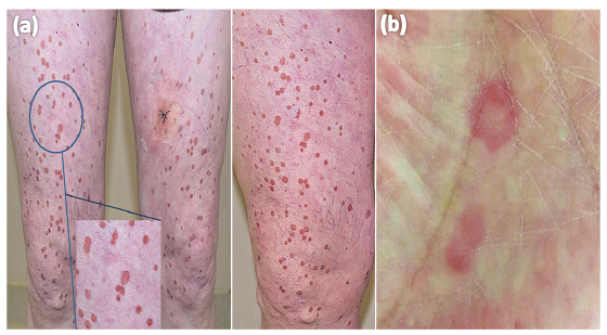 Click to show fullsize