


1Department of Dermatology and Allergology, Helsinki University Hospital and 2University of Helsinki, Meilahdentie 2, PO Box 160, FIN-00029 Helsinki, Finland. E-mail: anna.pankakoski@hus.fi
Accepted Feb 25, 2020; Epub ahead of print Mar 11, 2020
Acta Derm Venereol 2020; 100: adv00086
Juvenile bullous pemphigoid (BP) manifests clinically as blisters arising over an erythematous or urticarial base before the age of 18 years. Its diagnosis is based on similar criteria as in adult patients (1, 2). Some authors distinguish early childhood BP (3) from BP in adolescence, which is rarer (4). In recent years, the incidence of BP has increased in Finland (5), but the incidence of juvenile BP is unknown. To our knowledge, we report here the 3 first Nordic cases of juvenile BP. The main characteristics of the cases are summarized in Table SI.
Case 1. A 13-year-old Finnish girl with a history of atopia developed pruritic erythematous papules on the extremities. After 1 month, tense and flaccid blisters developed on her palms and soles (Fig. 1A, B). Direct immunofluorescence (DIF), circulating serum BP180 antibodies and elevated eosinophilia supported the diagnosis of BP. The blisters resolved after initiation of oral prednisolone (OP) of 60 mg/day (1 mg/kg/day). Adverse effects prompted tapering of systemic corticosteroids and discontinuation 3 months later. During tapering of prednisolone, oral methotrexate was initiated, at 15 mg/week, and showed a good response within 3 months. Six months after initiation of methotrexate BP180 antibodies were undetectable. One year after onset of BP the patient was still in remission and methotrexate could be discontinued.
Case 2. A 3-year-old boy with a history of iritis and juvenile polyarthritis and treatment with methotrexate, 5 mg/week, and infliximab, 100 mg every 8 weeks, presented with a sudden generalized eruption of erythematous maculo-papules on the face, limbs and trunk. Pruritic and tense over 1-cm sized blisters were formed on the limbs and soles (Fig. 1C, D). There was minor involvement of the oral mucosa. The patient had no fever or elevation of infection markers. Biopsy, DIF and circulating BP180 antibodies confirmed BP. OP was initiated at a dose of 30 mg (1.9 mg/kg/day) with methotrexate 10 mg/week, with a good response. OP was discontinued after 2.5 years of treatment. Corticodependence was observed when decreasing by between 5 and 10 mg/day with 3 relapses of BP. An exacerbation of polyarthritis further prolonged the long-term tapering of the drug. Dapsone 25 mg/day for 2 months was ineffective. Intravenous immunoglobulins (IVIg), 15 g every 2 weeks, induced remission, but was not sufficient to prevent new eruptions when methotrexate was paused and the dose of prednisolone further tapered, thus indicating the need for very slow corticosteroid tapering. One year and 4 months from starting IVIg, time intervals between the infusions were slowly increased from 2 to 16 weeks. Infliximab was paused when IVIg treatment was initiated, but reintroduced 3 years from onset of BP symptoms, and did not trigger a flare of BP. Six years after the diagnosis, BP180 antibody levels remain slightly elevated (13 U/ml), while the patient was still on methotrexate 10 mg/week, infliximab and IVIg. IVIg treatment has, thus far, continued for 6 years.
Case 3. A 3-month-old male infant presented with a sudden eruption of vesiculo-bullous lesions on the hands and soles, followed by widespread erythematous and urticarial plaques on the face, limbs and trunk. Serous bullae appeared later on the extremities mimicking clinically linear IgA disease (Fig 1E, F). The patient had received DTaP-IPV-Hib, rotavirus and pneumococcal conjugate vaccination 7 days before onset of skin symptoms. DIF and circulating BP180 antibodies confirmed BP diagnosis. Oral clarithromycin, 15 mg/kg/day, for 3 weeks in combination with topical low-potency corticosteroid treatment improved the skin symptoms. After 1 month, no new lesions appeared and by 4 months all lesions and pruritus had resolved. There were no signs of BP during the follow-up period of 8 months, by which time BP180 antibodies had decreased to 9 U/ml.
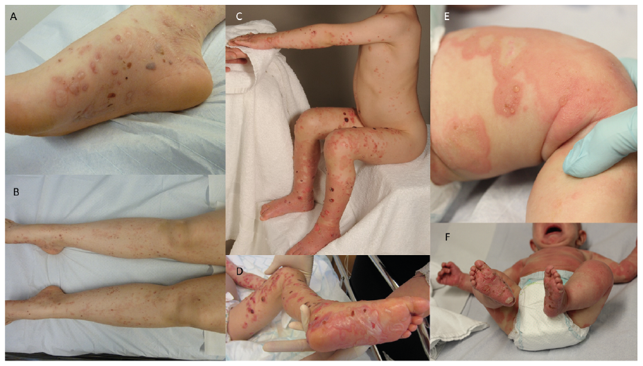
Fig. 1. Clinical photographs of patients with juvenile bullous pemphigoid (BP). (A, B) Case 1: Pruritic papular lesions on the extremities were followed by formation of serous and haemorrhagic bullae on the soles and medial arches of the feet. (C, D) Case 2: Widespread bullous and crusted lesions on erythematous base mainly affecting the skin of the extremities. (E, F) Case 3: Widespread erythematous plaques and annular lesions on the face, trunk and limbs alongside vesiculobullous lesions on all 4 extremities.
With fewer than 100 reported cases in the literature, juvenile BP is a rare acquired autoimmune disorder (6). The diagnostic features are similar for both paediatric and adult patients, and therefore juvenile BP should be seen as part of the same disease entity. However, the clinical hallmark of juvenile BP is acral distribution of skin lesions and frequent mucosal involvement, which are rare in adult patients with BP (7). Females are more frequently represented than males in the literature (62% vs. 38%) (3). Since BP and epidermolysis bullosa acquisita (EBA) display similar findings on DIF, Welfringer-Morin et al. (8) suggested using BP180, BP230, and anticollagen VII antibodies to differentiate both conditions. All of our cases presented high levels of circulating BP180 antibodies, which favoured pemphigoid rather than EBA.
There have been some reported cases of infliximab-induced BP (9). In case 2, the onset of BP was considered unrelated to infliximab. However, a similar cytokine profile of BP and juvenile arthritis might have been a predisposing factor for BP. In case 3, BP occurred one week after vaccination. In infantile BP onset of skin symptoms have been reported to occur within 2 weeks from vaccinations (10). Only larger studies could confirm whether the association is fortuitous. However, BP is not a contraindication to continue immunization.
Juvenile BP often runs a self-limiting course, with frequent spontaneous remission. Remission is reached within 1 year from disease onset and relapses are rare (11). Case 3 illustrates this typical course with a dramatic disease onset and rapid resolution of lesions with no following relapse. On the other hand, case 2 was challenging with a long duration of symptoms after diagnosis and still slightly elevated levels of BP180 antibodies several years later.
In adolescence, BP requires less hospitalization than other autoimmune bullous disorders (12). The rapid onset of widespread lesions and demanding topical treatment led to hospitalization in all 3 cases. Most commonly used treatments are oral corticosteroids and dapsone. Other successful treatment regimens include among others erythromycin, azathioprine, mycophenolate mofetil, IVIg and rituximab (13). Methotrexate has also showed good responses in some cases of juvenile BP (14). Two of our patients received methotrexate, case 1 as second-line treatment for BP and case 2 for polyarthritis prior to BP onset. In case 1 the response to methotrexate was rapid. It is likely that, in case 2, the onset of BP symptoms were somewhat postponed by methotrexate and triggered when the medication was paused. Based on these favourable experiences, methotrexate could be regarded as a possible treatment option in juvenile BP. After initial disease control the treatment should be based on the least aggressive therapy and lowest possible doses that prevent relapses.


 Click to show fullsize