


1Department of Dermatology and Allergology and 5Department of Plastic Surgery, Tampere University Hospital and Faculty of Medicine and Health Technology, Tampere University, Tampere, 2Department of Dermatology, Skin Cancer Unit, Helsinki University Central Hospital, Helsinki, 3Research, Development and Innovation Center, Tampere University Hospital and Health Sciences, Faculty of Social Sciences, Tampere University, 4Department of Pathology, Tampere University and Fimlab Laboratories, Tampere, Finland
Recognising patients with high risk cutaneous squamous cell carcinomas is essential in planning effective monitoring. The aim of this study was to determine the rate of local recurrences and metastases of cutaneous squamous cell carcinomas in a previously defined patient cohort in Finland. Pathology database search was performed to identify cutaneous squamous cell carcinoma patients and their medical records were reviewed. The cohort consisted of 774 patients with 1,131 cutaneous squamous cell carcinoma tumours. Overall, 4.2% (48/1,131) of the tumours were metastatic and 2.2% (25/1,131) had a local recurrence. Three of the metastatic tumours and 8 of the recurrent tumours had an invasion depth of ≤ 2 mm. The majority of metastases (28/48; 58%) were found within 3 months of the diagnosis of cutaneous squamous cell carcinoma. In conclusion, our study demonstrated recurrences and metastases even in the case of thin cutaneous squamous cell carcinomas and in high-risk cases close monitoring should be organised during the first years after diagnosis.
Key words: squamous cell carcinoma; keratinocyte carcinoma; metastasis; recurrent tumour.
Accepted Apr 2, 2020; Epub ahead of print Apr 7, 2020
Acta Derm Venereol 2020; 100: adv00121.
Corr: Niina Korhonen, Department of Dermatology and Allergology, Tampere University Hospital and Faculty of Medicine and Health Technology, Tampere University, FIN-33521 Tampere, Finland. E-mail: niina.korhonen@fimnet.fi
The majority of cutaneous squamous cell carcinomas can be cured with surgical excision but a small subset of these tumours metastasize. Our aim was to determine the rate of local recurrences and metastasis of these tumours in a patient cohort in Finland. Overall, 4.2% of the 1,131 tumours were metastatic and 2.2% of the tumours had a local recurrence. Over half of metastases were found within 3 months after tumour diagnosis. Patients with cutaneous squamous cell carcinomas should undergo individualised risk ascertainment and in high risk cases monitoring should be organised especially during the first years after diagnosis.
The incidence of cutaneous squamous cell carcinoma (cSCC) is increasing throughout the world (1). Although the vast majority of cSCCs can be cured with surgical excision, the rate of metastasis in cSCC has been estimated to range from 2% to 5% (2–5) and metastatic disease has a poor prognosis with a median survival of less than 2 years (2, 6). It is therefore important to recognise patients and tumours with high risk features early and effectively. We have previously reported trends in cSCC in a patient cohort in Finland over a 10-year study period (7). The objective of this present study was to determine the rate of local recurrences and metastases of cSCC in the same cohort and to analyse the histopathological features of the tumours together with patient characteristics, including any history of premalignant lesions, basal cell carcinoma (BCC) or cutaneous melanoma (CM).
A pathology database search was performed at Fimlab Laboratories to identify all patients in the Pirkanmaa region of Finland diagnosed with cSCC in 2006–2015. Details of these patients and their tumours were obtained by reviewing the clinical records of Tampere University Hospital. The data on the cSCC tumours included anatomic location, degree of differentiation and depth of invasion. Patients with multiple primary cSCC tumours diagnosed during the given period were included, and each tumour was analysed separately. The age of the patient at diagnosis of the primary tumour, gender and possible immunosuppression were recorded. The lifetime prevalence of actinic keratosis (AK), cSCC in situ, i.e. Bowen’s disease (BD), BCC and CM were ascertained for each patient from the medical records. The timing of the diagnosis of each local recurrence or metastasis of cSCC occurring within the period concerned was determined. In the case of multiple cSCCs, one primary tumour was designated as the recurrent or metastatic cSCC on the basis of the clinical notes. This retrospective study was approved by the institutional review board of Tampere University Hospital, Finland.
Categorical data were described in terms of the number of patients or number of tumours with percentages, and differences between such variables were tested with the Pearson chi-square test or Fisher’s exact test. All the analyses were conducted using IBM SPSS Statistics version 25.0 software (IBM SPSS, Chicago, IL, USA), and p-values under 0.05 were considered statistically significant.
Recurrent tumours
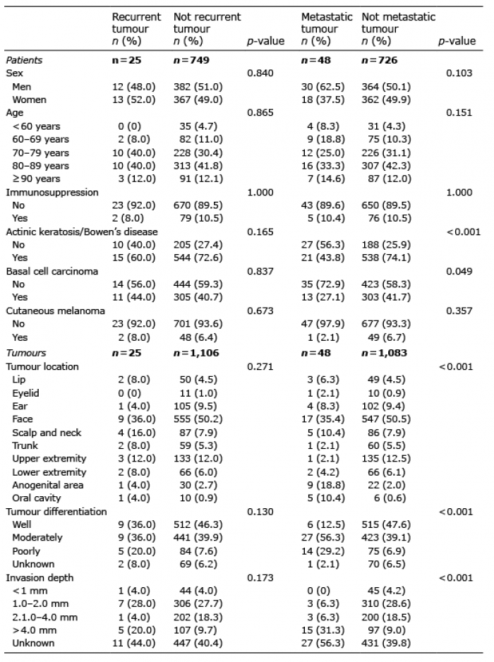
The cohort consisted of 774 patients with 1,131 cSCC tumours. Altogether, 25 of the 774 patients (3.2%) had a recurrent cSCC. Tumours in the location of the scalp and neck (4 recurrences in 91 tumours; 4.4% of cases in that location) or the trunk (2 recurrences in 61 tumours; 3.3%) showed more recurrences than would be expected in view of the overall recurrence rate (25 recurrences in 1,131 tumours; 2.2%), whereas only one recurrence was found in the case of the 106 tumours located on the ear (0.9%) (Table I). Nine out of the 25 recurrent tumours (36.0%) were well differentiated and 8 (32.0%) had an invasion depth of ≤ 2 mm (Table I). Five recurrent tumours had metastasized: 3 located on the face, one on the lip and one on the scalp. The median recurrence time was 13 months (range 2–72 months).
Table I. Characteristics of patients with recurrent cutaneous squamous cell carcinoma (cSCC) (n = 25; 3.2% of 774), recurrent cSCC tumours (n = 25; 2.2% of 1,131), patients with metastatic cSCC (n = 48; 6.2% of 774) and metastatic cSCC tumours (n = 48; 4.2% of 1,131)
Metastatic tumours
Of the 774 patients, 48 (6.2%) had a metastatic cSCC (Table I), including 21 patients (43.8%) who had a history of premalignant lesions, whereas 538 out of the 726 patients without any metastasis (74.1%) had had a premalignant lesion (p < 0.001). A total of 27.1% (13/48) of patients with a metastatic cSCC had BCC, compared with 41.7% (303/726) of those without any metastasis (p = 0.049). Any specific immunosuppression type (heart, kidney or liver transplantation, rheumatoid arthritis, chronic leukaemia or lymphoma) was not associated with meta-static cSCC. Altogether, 4.2% of the tumours (48/1,131) were metastatic, including 5 out of the 91 tumours on the scalp and neck (5.5%) but only one out of the 136 (0.7%) on an upper extremity (Table I). The majority of the metastatic tumours were moderately or poorly differentiated (41/48; 85.4%), and 3 (6.3%) had an invasion depth of ≤ 2 mm (Table I). The median time for the observation of a metastasis was 4 months (range 0–34 months), with 58% of them (28/48) found within 3 months of the cSCC diagnosis.
The proportion of the 1,131 cSCC tumours that were metastatic, 4.2%, is in line with previously reported metastasis rates of between 2% to 5% in cSCC (2–5). High-risk prognostic factors for cSCC include tumour location on the ear, lip or areas of long-lasting inflammation, a tumour diameter of > 2 cm, a histological depth of > 6 mm, moderately or poorly differentiated grade, certain histological subtypes (acantholytic, spindle, desmoplastic), perineural invasion, recurrence and immunosuppression (2). According to a recent meta-analysis, tumour depth is associated with the highest risk of local recurrence and metastasis in cases of cSCC (8). In our study, 6.3% of the metastatic tumours had an invasion depth of ≤ 2 mm, although tumours of that thickness have been considered to entail only a minimal risk (2). One previous study, for instance, states that no cSCC tumours ≤ 2 mm in thickness generated metastases (9).
The rate of local recurrences per tumour in the present series, 2.2%, is slightly lower than the figures of 2.7% (5) and 3.2% (10) reported previously. It should be remembered, however, that the recurrence rate depends on the subset of patients, the follow-up period and the treatment methods. In a systematic review, the pooled average of the local recurrences after standard surgical excision was 5.4% (11). Approximately one third of the recurrent tumours in our study were well differentiated and had an invasion depth of ≤ 2 mm, which are generally considered to be low-risk features (2). The risk of local recurrence has been reported to be dependent on increased tumour thickness (9, 12), tumour diameter (12) and desmoplasia (9).
Surprisingly many of the present scalp and neck cSCCs were recurrent and metastatic, and tumours on the trunk also showed more recurrences than was expected. On the other hand, recurrent tumours on the ear were rare. cSCCs located on the ears have been recognised as high-risk tumours (2, 3), while the scalp and neck have been identified as an intermediate-risk area and the trunk and limbs as a low-risk area (3). The status of cSCCs located on the trunk could be attributable to a lack of information, however, as these are infrequent compared with cSCCs of the head and neck. The guidelines contained in the 8th edition of the American Joint Committee on Cancer (AJCC) Staging Manual, for example, apply only to cSCCs of the head and neck (13). In addition, the wide diversity of cSCC histopathological subtypes could mean that the prognostic significance of some of them could be underestimated (13).
Where the majority of our metastases (58%) were found within 3 months of the diagnosis of cSCC, previous studies have reported that 73% of metastases occurred within the first year after resection (9) and all the metastases appeared within two years (14). There is no standardised follow-up schedule for patients with cSCC, but close monitoring has been recommended, particularly during the first years after diagnosis (2).
The fact that patients with metastatic cSCC had premalignant lesions and BCC less frequently than those who had non-metastatic cSCC suggests that patients without a history of precursor lesions may have an increased risk of developing biologically more aggressive cSCC tumours with a distinct aetiopathogenesis. On the other hand, patients previously treated for premalignant lesions or BCC may be subject to increased surveillance for new skin tumours and have their cSCC detected and treated early, before progression to a metastatic tumour.
In conclusion, our study demonstrated recurrences and metastases even in the case of thin cSCCs, with the majority of metastases being found within a few months of the cSCC diagnosis. Patients with cSCC should undergo individualised risk assessment and in high-risk cases close monitoring should be organised during the first years after diagnosis.
The study was supported financially by the Finnish Dermatological Society (grant awarded to Dr. Korhonen). This organisation had no involvement in the design and conduct of the study, the collection, management, analysis or interpretation of the data, the preparation, review, or approval of the manuscript, or the decision to submit the manuscript for publication.
The authors have no conflicts of interest to declare.


 Click to show fullsize