


1Dr. Phillip Frost Department of Dermatology and Cutaneous Surgery, Miami Itch Center, Miller School of Medicine, University of Miami, Miami, FL, USA, 2Department of Integrative Physiology, and 3Department of System Neuroscience, National Institute for Physiological Sciences, Okazaki, Japan
Itch is an unpleasant and aversive somatosensory experience. These negative emotions significantly affect mental health in patients with chronic itch; it is therefore important to understand the brain mechanism of negative emotions due to itch. The amygdala is a key hub of networks regulating negative emotions due to itch. However, the exact network involved in this process is unknown. This study used functional magnetic resonance imaging to investigate the amygdala network processing itch in 25 healthy subjects. Brain activity was measured during electrical itch stimuli using functional magnetic resonance imaging. During itch stimuli the amygdala exhibited increased functional connectivity with key brain regions of the serotonergic system responsible for negative emotions (the medial habenula and the median raphe nucleus) and with the memory system, which is responsible for consolidating emotional experiences (the parahippocampus and perirhinal cortex). The serotonergic and memory systems may become therapeutic targets to prevent or reduce diminished mental health commonly seen in chronic itch patients.
Key words: itch; negative emotion; serotonergic system; memory system.
Accepted Nov 4, 2020; Epub ahead of print Nov 25, 2020
Acta Derm Venereol 2020; 100: adv00345.
doi: 10.2340/00015555-3703
Corr: Hideki Mochizuki, Dr. Phillip Frost Department of Dermatology and Cutaneous Surgery, Miami Itch Center, Miller School of Medicine, University of Miami, 1600 NW 10th Ave, Miami, FL, 33136, USA. E-mail: hxm414@miami.edu
Itch is an unpleasant somatosensory experience, which negatively affects mental health in patients with chronic itch. This study found that itch activates key brain regions of the serotonergic system, responsible for negative emotions, and the memory system, responsible for consolidating emotional experiences. This finding provides an important insight into the mechanism of how itch can influence mental health. The serotonergic and memory systems may become therapeutic targets to prevent or reduce diminished mental health commonly seen in chronic itch patients.
Itch is an unpleasant and aversive somatosensory experience. These negative emotions significantly affect mental health in patients with chronic itch, which can lead to the development of chronic stress, depression, and anxiety disorder (1, 2). These psychological co-morbidities are common in patients with chronic itch (1–3). Therefore, understanding the neural mechanism behind negative emotions due to itch is necessary to prevent and treat psychological comorbidities due to itch.
The amygdala is a key hub of networks for processing negative emotions (4–8). Electrical stimuli to the amygdala can provoke various negative emotions, including fear, disgust, sadness, stress and anxiety (4). Thus, amygdala networks are foundations of emotional experiences. A recent animal study found neurones responsive to itch stimuli in the amygdala, such as the central nucleus of the amygdala (CeA) and basolateral amygdala (BLA) (9). Moreover, this study demonstrated that depression/anxiety-like behaviour is influenced by the manipulation of activity in itch-responsive amygdala neurones. These findings indicate that an amygdala network for processing itch plays a crucial role in the regulation of negative emotions due to itch. However, it largely remains unknown what functional network the amygdala constitutes for processing itch. The aim of this study was to investigate the itch-related amygdala network in the brains of healthy people, using functional magnetic resonance imaging (fMRI).
Subjects
The present study analyzed fMRI data and itch rating data obtained from our previous study. In the previous study, we investigated a key network responsible for itch perception, focusing on the posterior insular cortex (10). On the other hand, the present study focused on the amygdala, a key brain region of negative emotions due to itch, and investigated what network the amygdala constitutes for processing itch. The current fMRI study was conducted with 25 healthy Japanese subjects (7 females and 18 males), with a mean ± standard deviation (SD) age of 28 ± 9 years. Written informed consent was obtained from all subjects. The study complied with the principles of the Declaration of Helsinki, and was approved (Approved number: 12A014) by the Ethics Committee of the National Institute for Physiological Science (Japan).
Itch stimuli
Electrical itch stimuli (current 0.35 mA, frequency 50 Hz, pulse width 10 ms, 125 repetitions of pulses) were applied to the left wrist to provoke an itch sensation. A session comprised 6 blocks, in which a short duration of electrical stimulus (2.5 s) was applied 5 times at 5-s intervals. The interval between blocks ranged from 62.5 to 82.5 s. At the end of the session subjects were asked to rate the mean itch sensation of 6 blocks using a numerical rating scale (NRS), ranging from 0 (no itch) to 10 (the worst itch).
Magnetic resonance imaging measurement
Brain activity during the session was measured using a 3 Tesla magnetic resonance imaging (3-T MRI) scanner (Allegra, Siemens, Erlangen, Germany). For functional imaging, 156 volumes of T2*-weighted, gradient-echo, echo-planar imaging (EPI) sequences, 39 transaxial slices, thickness 3.0 mm, repetition time (TR) 2,500 ms, echo time (TE) 30 ms, flip angle (FA) 80°, field of view (FOV) 192 mm, and matrix 64 × 64. The fMRI data were pre-processed (realignment, spatial normalization and smoothing) and analysed using statistical parametric mapping software (SPM8; The Wellcome Trust Centre for Neuroimaging, London, UK).
Identification of itch-related activation inside the amygdala
We searched a location showing significant activation due to itch within the amygdala. Thus, the statical test was restricted within the amygdala. First, uncorrected p = 0.001 was applied as a statistical threshold to identify increased activity in brain regions during itch stimuli. Brain regions outside the bilateral amygdala were filtered out using a mask image of the amygdala (Wake Forest University (WFU) PickAtlas; https://school.wakehealth.edu/Research/Labs/Radiology-Informatics-and-Image-Processing-Laboratory/Software-Development#PickAtlas). Small volume correction (SVC) was then applied using the mask image, to determine whether activations inside the amygdala were statistically significant (statistical threshold: family-wise error rate (FWE) p < 0.05). The location with the highest z-score was used as a seed brain region for subsequent functional connectivity analysis.
Functional connectivity analysis
Psychophysiological interaction (PPI) analysis implemented in SPM (11) was used to identify brain regions that increased functional connectivity with the seed brain region during itch stimuli. The PPI was calculated using the time series of the blood oxygenation level-dependent (BOLD) signal within a 3-mm radius sphere around the coordinate of the seed brain region. The interaction term was subsequently reconvolved with the haemodynamic response function. The reconvolved interaction term was then entered as a regressor in a first-level model, together with the time series of the seed region and the psychological vector of interest (i.e. regressor used to identify the brain region activated by itch stimuli). Then, the models were estimated (first-level analysis). Subsequently, a second-level random effects analysis was performed. Brain regions that showed significantly increased or decreased connectivity with the seed region during itch stimuli were identified. In addition, this study examined whether the intensity of functional connectivity with the seed brain region correlated with itch NRS. Statistical threshold for the above analyses was set at p < 0.05 for intensity after the whole-brain FWE correction.
Activation inside the amygdala due to itch stimuli
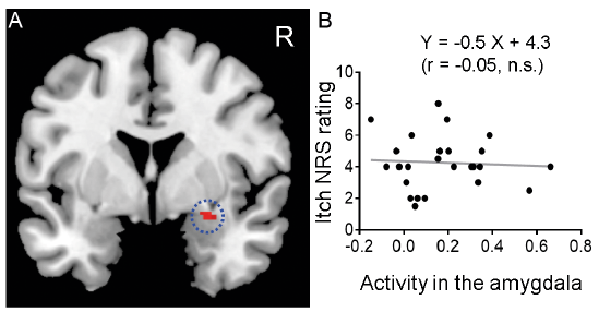
Mean ± SD itch itch NRS rating of all subjects collected at the end of the session was 4.3 ± 1.7. Significant activation of the right amygdala was observed (Fig. 1a). There was no significant correlation between itch-related activity in the amygdala and perceived itch sensation (Fig. 1b).
Fig. 1. The amygdala. (a) Significant activation due to itch in the contralateral (right) amygdala (MNI coordinates: 28, 2, –14, z-score: 3.75). (b) Correlation between itch numerical rating scale (NRS) rating and activity in the amygdala during itch stimuli.
Psychophysiological interaction analysis
During itch stimuli there was a significant increase in functional connectivity between the amygdala and the temporal cortex, including the perirhinal cortex and parahippocampus, ventromedial prefrontal cortex (vmPFC), midcingulate cortex (MCC), supplementary motor area (SMA), basal ganglia (BG) (mainly the globus pallidus), middle insula and cerebellum (Fig. 2 and Table I). There was also an increase in functional connectivity between the amygdala and the epithalamic regions, including the left habenula (the habenula is located adjacent to the posterior commissure (PC)) (12–17) (Fig. 2 and Table I). This result is consistent with a previous animal study that demonstrated that neural activity in the habenula is significantly increased by histamine-induced itch (18, 19).
Fig. 2. Functional connectivity with the amygdala. Brain regions that increased functional connectivity with the amygdala during itch stimuli. SMA: supplementary motor area; MCC: midcingulate cortex; vmPFC: ventromedial prefrontal cortex. Red arrows: habenula. Blue arrows: posterior commissure (PC).
Table I. Functional connectivity with the amygdala during itch stimuli
The habenula regulates emotions and motivations by interacting with serotonergic neurones in the raphe nuclei and dopaminergic neurones in the midbrain (12–14, 19). Further PPI analysis was therefore conducted to investigate whether activity in the left habenula increased functional connectivity with these brain regions during itch stimuli. During itch stimuli the left habenula increased functional connectivity with the raphe nucleus (MNI coordinates: 2, –28, –24, z-score: 5.02) (Fig. 3). In particular, the MNI coordinates were localized in the median raphe nucleus (MRN), as determined in a previous human study using positron emission tomography and serotonin ligand (20).
Fig. 3. Functional connectivity with the habenula. To investigate whether the brainstem increases functional connectivity with the left habenula during itch stimuli, an additional psychophysiological interaction (PPI) analysis was conducted. The PPI was calculated using the time series of the BOLD signal within a 3-mm radius sphere around the coordinate of the left habenula (Table I). The statistical threshold for this analysis was set at p < 0.05 with the whole-brain family-wise error rate (FWE) correction. Significant functional coupling was observed between the habenula and the raphe nucleus. For visual purposes, the functional brain image was thresholded with uncorrected p < 0.001.
This study identified brain regions that comprise a functional network with the amygdala for processing itch, including key brain regions associated with provocations and regulations of emotions, such as the MRN, habenula and vmPFC, and key brain regions of memory consolidation, such as the perirhinal cortex and parahippocampus. These brain regions may become important therapeutic targets for reducing and preventing psychological comorbidities due to itch.
Amygdala
The amygdala was significantly activated by itch stimuli. A previous animal study reported that both CeA and BLA were activated by itch stimuli (9). In accordance with this finding, the activity in the amygdala observed in the current study was adjacent to these 2 nuclei (21). Sanders et al. (9) discussed that the majority of itch-responsive amygdala neurones are associated with general aversion due to an experience of itch, while some neurones with specific response to itch are associated with itch-related scratching behaviour. The present study, did not find a significant correlation between itch-related activity in the amygdala and NRS rating of itch. These results suggest that the significant activation of the amygdala observed in the current study may be associated with general aversion accompanied by itch rather than itch intensity.
Habenula, median raphe nucleus, and ventromedial prefrontal cortex
Several studies have investigated the precise location of the habenula in humans and monkeys, using MRI, postmortem brains, and immunohistochemical approaches (12, 15–17). These studies have commonly reported that the habenula is located adjacent to the PC. In accordance with this anatomical feature, the current study observed that a brain region adjacent to the PC showed increased functional connectivity with the amygdala during itch stimuli (Fig. 2). The habenula is composed of the lateral and medial habenula. A previous human study using high-resolution MRI, such as 7T-MRI, has demonstrated that the medial habenula is located more anterior than the PC in a horizontal brain section, while the lateral habenula and the PC are in the same location (17). This study has also demonstrated that the height of the medial habenula is the same as, or slightly more dorsal to, the PC in a coronal brain section, while the lateral habenula is located more dorsal to the medial habenula. The same result was also reported in a previous immunohistochemical study in monkeys (12). These anatomical features of the medial habenula were also seen in the left habenula in the current study (Fig. 2). In addition, an animal study has demonstrated that the medial habenula is significantly activated by itch stimuli (18), supporting the current finding.
The current study observed that functional connectivity between the medial habenula and the MRN increased during itch stimuli. This is consistent with the finding that the medial habenula sends projections to the MRN via the interpeduncular nucleus (20). The raphe nuclei, including the MRN and dorsal raphe nucleus (dRN), are rich in serotonergic neurones. Previous studies have demonstrated that serotonin release from the MRN, but not dRN, is associated with anxiety, and low dRN/MRN serotonergic activity ratio is associated with depression (5, 22–24). A previous study reported that light-induced activation of itch-responsive amygdala neurones enhanced depression/anxiety-like behaviour due to itch (9). Itch-induced increment of the habenula-MRN functional coupling may enhance serotonin release. Future studies should address this question.
The functional coupling of the vmPFC and amygdala plays an important role in emotional control (25–27). It has also been reported that patients with lesions in the vmPFC have difficulty in controlling emotional reactions, resulting in emotional outbursts and impulsive behaviours (28). An optogenetic mice study reported that both amygdala and vmPFC were activated by itch stimuli, and that light-induced manipulation of activity in the itch responsive amygdala neurones influenced depression/anxiety-like behaviour due to itch (9). It is possible that a broad functional network, involving the vmPFC, amygdala, habenula and MRN, may regulate negative emotions due to itch.
Temporal cortex
The significant increment in functional connectivity between the amygdala and temporal cortex including the perirhinal cortex and parahippocampus may also contribute to negative emotions of itch. The temporal cortex is a key structure of the memory system. In particular, the perirhinal cortex and parahippocampus provide the major source of cortical input to the hippocampal formation (29). Lesions in these brain regions cause severe memory impairment (30). The perirhinal cortex receives neural signals from the amygdala (31). This communication induces plastic changes in the memory system, resulting in prioritizing an event in the memory (32–35). Experiences of itch, including negative emotions, may be consolidated in memory through the network involving the amygdala and temporal cortex. This consolidation may make negative emotions due to itch last longer, which could facilitate or cause diminished mental health in patients with chronic itch.
In pain studies, several lines of evidence have demonstrated that pain memories affect the subjective sensation of pain. For example, both mice and human subjects became hypersensitive to pain when tested in an environment previously associated with an aversive tonic pain experience (36). Clinical studies have reported that patients with chronic pain were suddenly relieved from chronic pain after amnesia (memory loss) (37, 38). In itch studies, it has been demonstrated that merely thinking about itch can cause itch perception in patients with chronic itch, which rarely happens in healthy subjects (39). Itch memory may be an important factor in determining the subjective perceived intensity of acute and chronic itch (40).
Conclusion
This study used fMRI to identify brain regions involved in an itch-related amygdala network in humans. These brain regions are involved in the serotonergic system and memory system. This suggests that negative emotions due to itch may be associated with the serotonergic system, and the emotional experience of itch may be consolidated in the brain through the memory system. Future studies focusing on these systems may lead to the development of novel treatments to prevent and reduce psychological comorbidities due to itch, which are commonly seen in patients with chronic itch.
This study was partly supported by a grant from the Center for Novel Science Initiative.
The authors have no conflicts of interest to declare.


 Click to show fullsize
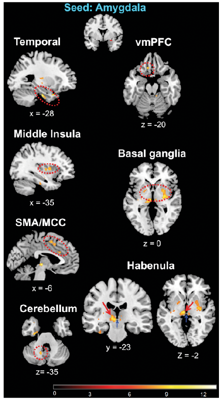 Click to show fullsize
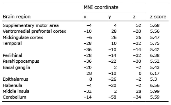 Click to show fullsize
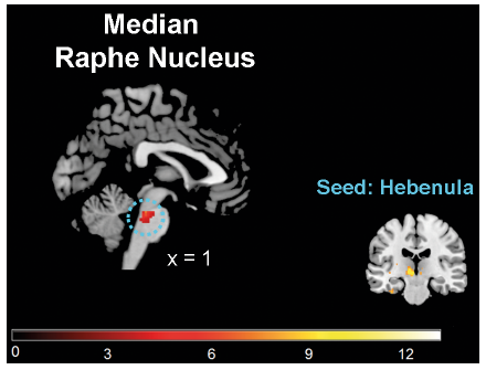 Click to show fullsize