


1Department of Dermatology, Institut Clínic de Medicina i Dermatologia, 2GVHD and Long-Term Follow-up Unit, Hematopoietic Stem Cell Transplantation Unit, Department of Hematology, Institut Clínic de Malalties Hemato-Oncològiques, Hospital Clínic, University of Barcelona, Barcelona, Spain and 3Institut d’Investigacions Biomèdiques August Pi i Sunyer, Barcelona, Spain
Sclerodermoid chronic graft-versus-host disease (scGVHD) is a severe complication of allogeneic haema-topoietic stem cell transplantation. The aim of this study was to investigate the usefulness of high-frequency ultrasound of the skin in assessing the inflammatory patterns and prognosis of patients with scGVHD. A prospective study was carried out with patients who developed scGVHD in the period June 2016 to April 2018. Clinical and ultrasound examinations were performed on the first visit and at 6-month follow-up. A total of 24 patients were included in the study. A 6-month follow-up high-frequency ultrasound of the skin was performed on 20 of the 24 patients. Abnormal B-mode findings in high-frequency ultrasound of the skin consisted of hypoechogenic dermis, hypoechogenicity of septa and hyperechogenicity of lobules in hypodermis. No differences were observed in these basal parameters between treatment progressive/non-responding and inactive/responding scGVHD groups of patients. Basal Doppler showing increased vascular flow with a systolic peak ≥10 cm/s and a vascular resistance index ≥ 0.70 was observed only in those patients who developed progressive/non- responding scGVHD (62.5% vs 0% p = 0.006). In conclusion, Doppler ultrasound is a useful tool to assess the inflammatory activity and outcome of scGVHD. These findings could enhance patient management and help to guide treatment decisions.
Key words: graft-versus-host disease; Doppler ultrasound; diagnosis; sclerodermoid; inflammation; allogeneic haematopoietic stem cell transplantation.
Accepted Jan 19, 2021; Epub ahead of print Jan 21, 2021
Acta Derm Venereol 2021; 101: adv00395.
doi: 10.2340/00015555-3747
Corr: Priscila Giavedoni, Department of Dermatology, Hospital Clínic de Barcelona, Universitat de Barcelona, Villarroel 170, ES-08036, Barcelona, Spain. E-mail: pgiavedo@clinic.cat
Graft-versus-host disease is one of the most serious complications of haematopoietic stem cell transplantation. However, treatment with immunosuppressants to reduce inflammation increases the risk of recurrence of the neo-plasm that necessitated the stem cell transplantation. Hence, it is extremely important in these patients to determine whether graft-versus-host disease is active, in order to make appropriate treatment decisions. High-frequency skin ultrasound is an imaging technique that is used increasingly in dermatology and can be useful in diagnosing this disease. Skin inflammation can be quantified using the colour Doppler mode. Clinical and ultrasound monitoring enable objective assessment of these patients.
Chronic graft-versus-host disease (cGVHD) remains a significant complication in patients undergoing allogeneic haematopoietic stem cell transplantation (allo-HSCT) and is a leading cause of late morbidity and mortality (1). cGVHD has a wide range of pleomorphic manifestations, and many complications can emerge from both the disease and its treatment (2). In 2005, the National Institutes of Health (NIH) cGVHD Consensus Conference Criteria developed guidelines for diagnosis, grading organ involvement and overall disease severity, as well as indications for treatment and recommendations for monitoring disease progression and response to therapy, which were revised in 2014 (3, 4). However, these guidelines are based solely on expert opinion, and require validation through further research.
Skin is the most frequently affected organ in cGVHD, and manifestations are highly variable. According to the NIH cGVHD Consensus Conference, diagnostic clinical features include poikiloderma, lichen planus-like eruption, deep sclerotic features, morphea-like superficial sclerotic features, and lichen sclerosus-like lesions. The sclerodermoid form of cGVHD (scGVHD) often progresses from morphea-like plaques to generalized scleroderma and fasciitis. resulting in joint contractures and severe functional disability (4).
One of the most challenging aspects of the diagnosis and management of scGVHD is the differentiation be-tween reversible symptoms related to active cGVHD and non-reversible symptoms related to residual permanent damage, such as fibrosis. Although several candidate biomarkers of cGVHD inflammatory activity have been proposed, to date none of them has been validated. Therefore, there is a need for the development of more quantifiable and reproducible measurement tools, which can be used to guide clinical decisions for patients with scGVHD.
High-frequency ultrasound of the skin (HFUS) is a technique that is used increasingly in dermatology for the diagnosis and evaluation of benign and malignant disorders, infectious diseases, and inflammatory diseases, including systemic sclerosis, localized morphea, and lichen sclerosus (5–7). HFUS with colour Doppler has shown great promise in the diagnosis of systemic sclerosis through the analysis of structural changes in tissue and vascularity (8). It has also been suggested that colour Doppler may prove useful in monitoring disease activity and response to treatment, and may be more sensitive than clinical assessment alone in detecting the oedematous phase that occurs before the fibrotic stage (9, 10). The usefulness of elastography to measure the rigidity of patients with sclerosing skin diseases has been described. Studies have also been performed with HFUS to measure skin thickness in these patients (11–13).
To the best of our knowledge, there is no information on the use of colour Doppler ultrasound in the evaluation of inflammatory activity in patients with scGVHD. The aim of this study was therefore to evaluate the usefulness of HFUS with colour Doppler to determine the inflammatory activity of scGVHD.
Patients
Allo-HSCT recipients who had developed scGVHD between 1 June 2016 and 30 April 2018 were enrolled in this study. All patients were assessed by an expert panel of dermatologists and haematologists, working in a multidisciplinary GVHD group. The study was conducted according to the ethical principles of the Declaration of Helsinki, and in accordance with the International Conference on Harmonization Guideline for Good Clinical Practice. The protocol was approved by the ethics committee of the Hospital Clínic of Barcelona, Spain (HCB/2017/0599). All patients provided written informed consent before taking part in the study.
Skin, joint and fascia involvement by cGVHD were diagnosed according to the 2014 NIH cGVHD diagnostic criteria. Limitation of mobility due to inflammation of the fascia and/or rigidity of the joints was measured by the Photographic Range of Motion (P-ROM) scale. In each patient, the percentage of body surface area (BSA) affected by sclerosis changes was evaluated. Data regarding systemic immunosuppression treatment for scGVHD at the time of study enrolment and at 6-month follow-up were also recorded.
Skin ultrasound
HFUS was performed by a dermatologist trained in the diagnosis and management of cGVHD and in HFUS. In Department of Dermatology, Hospital Clínic Barcelona, we have a specific consultation to perform the HFUS. Patients are scheduled to allow 1 h to perform each HFUS. The HFUS was performed in the same week as the dermatological evaluation.
Esaote MyLab™ClassC equipment (ESAOTE ESPAÑA, Barcelona, Spain) was used with high-frequency probes between 10, 18 and 22 MHz. The software is based on Windows® XP, and uses Digital Imaging and Communications in Medicine (DICOM) services to download worklists and save acquired ultrasound images on a network storage recordable compact disc (CD-R), DVD, or a removable device connected to a USB, and to print images on a network copy device.
The entire body surface of each subject was examined, and ultrasound examination was divided into 7 anatomical sites (face, neck, upper and lower extremities, trunk, abdomen, and pubis). Between 5 and 10 images were taken at each anatomical site. The anatomical sites were not divided into segments. Each area was studied over at least 50% of the surface; the areas with most inflammation were recorded for comparison at follow-up. The minimum time to perform the HFUS was 30 min, and the maximum was 60 min.
HFUS was performed using both B-mode grey-scale ultrasound imaging and colour Doppler mode. The colour Doppler mode was used to determine the direction of blood flow. Power Doppler mode was used to assess skin vascularization, as it is more sensitive to low-flow/low-speed vessels found in inflammatory skin lesions. Spectral Doppler analysis was performed to distinguish veins from arteries, and, in the latter, to determine the systolic peak, diastolic peak, and resistance index. Doppler measurements were made with an insonation angle ≤ 60°. The pulse repetition frequency was fixed at 750. The colour gain was variable and adjusted to the value immediately below the noise threshold.
The skin was considered to have normal ultrastructure if the epidermis measured up to 0.1 mm (14) and was seen as a thin hyperechogenic line, the normal dermis is seen as a hyperechoic band, and the hypodermis was seen with hypoechoic areas corresponding to the lobules, with thin hyperechoic bands corresponding to the septa (15). Normal fascia was defined as a hyperechoic band, and normal muscle showed a fibrillar pattern with narrow hyperechoic grouped bands (Fig. 1a) (16).
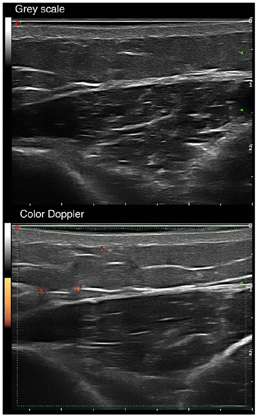
Fig. 1. High-frequency ultrasound of normal skin. The upper image shows hypoechogenicity of the dermis, loss of dermo-hypodermal differentiation, increased hypoechogenicity and thickness of the septa, with hyperechogenicity of the lobules. The lower image shows the increased flow in color Doppler mode.
Using current equipment as in this study, blood flow is rarely detected in the normal dermis on colour Doppler, and isolated vessels smaller than 1 mm in diameter are identified in the hypodermis (14). Arterial vessels in healthy skin have a low velocity, with a systolic peak of less than 10 cm/s with spectral Doppler (Fig. 1b) (14). Therefore, in this study, the following findings were described as negative Doppler: isolated vessels in the dermis and hypodermis, with a systolic peak of less than 10 cm/s and a resistance index of less than 0.7 hertz.
The variables analysed in the HFUS B-mode considered indicative of active disease were those normally described in fibrous skin diseases: hypoechogenic dermis, deletion of the dermo–epidermal junction, hypoechogenicity of the septa and hyperechogenicity of the lobes in the hypodermis, hypoechogenic fascia, and/or loss of the muscular fibrillar pattern (14, 17, 18). Affected areas were compared with perilesional or contralateral healthy skin, making it possible to determine whether there was an increase or decrease in echogenicity.
The variables of Doppler mode indicative of inflammatory disease were vessels greater than 1 mm in diameter in the dermis or hypodermis, and arterial vessels with a high velocity flow (systolic peaks > 10 cm/s and vascular resistance index > 0.70 hertz). These cut-off values were set based on those published to date (14, 17, 19, 20), and higher values than those previously described were used to increase the specificity of the test.
Study design
Clinical and ultrasound examinations were performed when patients were first seen in dermatology and at 6-month follow-up. Patients were classified into 3 groups according to their clinical features: (i) patients with clinical inflammatory activity of scGVHD presenting signs and symptoms of morphea-like plaques (oedema, erythema, hardening, orange peel appearance, and pain on palpation in the previous 3 months); (ii) patients with clinical inflammatory activity of scGVHD presenting signs and symptoms of fasciitis (pain, retraction of skin and muscles, and limited mobility); and (iii) patients with chronic changes of sclerosis (hardening of the skin and limitation of movement), but without clinical signs of inflammation, that were classified as clinically inactive. Patients not fulfilling these criteria were considered as indeterminate. The patients were divided into 2 groups according to their clinical evolution at 6-month follow-up. The first group (progressive/non-responding scGVHD) consisted of patients who reported clinical worsening of inflammatory and/or sclerodermic signs or symptoms (oedema, erythema, pigmentation changes, hardening, orange peel appearance, pain on palpation and or limitation of mobility), and who required treatment with systemic corticosteroids and/or a new immunosuppressive therapy line for cutaneous scGVHD, or those in whom stopping/tapering of systemic corticosteroids was not possible due to skin disease activity. The second group (inactive/responding scGVHD) consisted of patients reporting clinical improvement, or in whom changes were categorized as residual (non-inflammatory hardening and limitation of mobility unmodified within the 6 months), and those in whom systemic immunosuppression treatment could be reduced or stopped. Patients who did not need to start immunosuppressive therapy within the period of follow-up were also included in this second group.
Colour Doppler ultrasound was performed when patients were first seen and on follow-up at 6 months. The study was repeated on the same areas and with the same parameters as in B-mode and Doppler colour.
Statistical analysis
Fisher’s exact test was used to compare categorical variables. Sensitivity, specificity, positive predictive value (PPV), and negative predictive value (NPV) were calculated for each categorical variable that showed significance after Fisher’s exact test. p-values < 0.05 were considered significant. Statistical analyses were performed using the computing environment R and RStudio.
Characteristics and outcome of sclerodermoid chronic graft-versus-host disease
Characteristics of patients and GVHD are summarized in Table I. Fourteen patients were considered to have clinically inflammatory disease, 2 clinically inactive disease, and 8 indeterminate disease (Fig. 2). A total of 7 patients (6 in the clinically active group and one in the indeterminate group) were receiving prednisone, at a dose of > 10 mg/day. Clinically inactive patients were not receiving prednisone at that time.
At 6 months of follow-up, 8 patients were classified as having treatment progressive/non-responding scGVHD, and 16 patients had responding scGVHD (Fig. 2).
Table I. Clinical characteristics of patients with sclerodermoid chronic graft-versus-host-disease (scGVHD)
Fig. 2. Clinical status of patients and Doppler ultrasound (DU) findings at the beginning of the study and at 6 months of follow-up. DU+: Doppler ultrasound positive (systolic peaks ≥ 10 cm/s and vascular resistance index ≥ 0.70 hertz). DU– : Doppler ultrasound negative (systolic peaks <10 cm/s and vascular resistance index < 0.70 hertz).
Ultrasound results
At first evaluation, abnormal B-mode findings in HFUS were frequent and consisted of hypoechogenic dermis (n = 12/24, 50%), hypoechogenicity of septa and/or hyperechogenicity of lobules in the hypodermis (n = 8, 33.3%), hypoechogenic fascia (n = 8, 33.3%), and myositis (n = 3, 12.5%) (Table II, Fig. 3a). No differences were observed in these basal parameters between treatment progressive/non-responding and responding scGVHD groups of patients.
Table II. Baseline sonographic characteristics of patients with sclerodermoid chronic graft-versus-host-disease (scGVHD)
Fig. 3. High-frequency ultrasound of skin in a patient with sclerodermoid chronic graft-versus-host-disease (scGVHD). Upper image: B-mode; lower image .
Abnormal findings in Doppler mode consisted of the presence of vessels in the dermis in 5/24 (20.8%) patients, vessels at the dermo-epidermal junction in 12/24 (50%), and vessels in the hypodermis in 10/24 (58.3%). Basal Doppler mode showing increased vascular flow with a systolic peak ≥ 10 cm/s and vascular resistance index ≥ 0.70 hertz was observed only in those patients who developed progressive/non-responding scGVHD (62.5% vs 0% in the inactive/responding group, p = 0.006) (Table II) (Fig. 3b). Clinically these patients showed persistence of cutaneous inflammatory lesions and continued to require steroids or immunosuppressive drugs to treat scGVHD. Moreover, 81% of inactive/responding patients had a negative basal Doppler. Thus, basal Doppler ultrasound findings had a PPV of 100% (95% confidence interval (95% CI) 69–100) with 100% specificity (95% CI 69–100). Sensitivity was 62% (95% CI 35–85) and the NPV was 57% (95% CI 29–82).
In 20 patients, an ultrasound was also performed at 6 months of follow-up (Fig. 2). A correlation was observed between Doppler findings and clinical status of scGVHD. Thus, all patients with vascular signs of inflammation by Doppler mode (n = 7) had progressive/non-responding disease. In contrast, those with normal Doppler ultrasound (n = 13), except one, had responding scGVHD. Moreover, normalization of abnormal findings of Doppler was observed in all patients who presented inflammatory signs at study entry and who were clinically categorized as responders at follow-up.
No correlation was found between P-ROM scale and clinical inflammatory activity of scGVHD: 12 patients from the group of progressive/non-responding scGVHD (75%) and 5 (62.5%) from the inactive/responding group presented abnormal P-ROM scale results (kappa correlation coefficient 0.40).
To the best of our knowledge, this is the first study to analyse the usefulness of Doppler ultrasound in the assessment of patients with scGVHD. A statistically significant correlation was found between basal Doppler mode findings indicating inflammatory activity and clinical outcome. The Doppler findings found to correlate with inflammation in patients with scGVHD were a systolic peak > 10 cm/s and a resistance index > 0.7 hertz.
HFUS has the following advantages: it is an inexpensive, simple, and non-invasive tool, which does not use ionizing radiation. In addition, HFUS can be performed serially during dermatological appointments, and there is a low intra- and inter-observer variability (9, 18). The main drawback of HFUS is that it requires significant operator skills and training, and it is time-consuming. Several studies have shown that HFUS is able to measure skin thickness in patients with systemic sclerosis (10, 18, 21). Other studies have also shown that HFUS findings can vary according to the phase of systemic sclerosis and morphea (17). HFUS can thus identify the oedematous phase, which may precede palpable skin involvement in patients with systemic scleroderma with short disease duration, and can reflect disease severity. Wortsman et al. (17) reported that, in patients with morphea, skin echogenicity characteristics during the active inflammatory phase of the disease were different from those of the atrophic phase. In that study, the most accurate sonographic signs of lesion activity were observed with Doppler mode, which showed increased subcutaneous tissue echogenicity and increased cutaneous blood flow (both 100% sensitivity and specificity).
Gottlöber et al. (13) were the first to describe the usefulness of HFUS in scGVHD, but in their study Doppler mode examination was not used. Five patients were studied, and HFUS evidence of skin thickness decrease was observed after treatment. In another study, 5 patients with scGVHD were evaluated using 20 MHz HFUS before and after therapy. The authors reported only small reductions in skin thickness after treatment (12). In the present study, HFUS was performed using high-frequency probes between 3 and 22 MHz, and abnormal B-mode findings were observed in most patients No correlation was observed between these findings and clinical outcome. However, changes in Doppler mode appear to be useful in diagnosing inflammation in these patients, employing a non-invasive technique.
Currently the diagnosis of scGVHD and treatment response evaluation relies on clinical examination only. According to the 2014 NIH Consensus Conference, response is measured using the NIH Skin Score, NIH Joint and Fascia score and P-ROM (4). These scores are based on clinician assessments of BSA affected, grade of impaired mobility, contractures, impact on functional status, and semi-quantitative scales for assessing clinician and patient-perceived severity of sclerosis. Response is defined according to changes in these scores before and after therapy. In comparison with other scales, such as the Vienna Skin Score or the Johns Hopkins sclerosis and fasciitis scales, the NIH Skin Score was reported to be the only one that correlated with both clinician and patient perception of improvement or worsening and with overall survival (22). However, the heterogeneity of skin manifestations included into the NIH Skin Score and the variability in the intra- and inter-observer are both limitations for its use in daily clinical practice. Furthermore, sclerotic changes take many months to resolve and NIH score may not be sensitive enough to detect slow, but meaningful, changes resulting from treatment, or to differentiate between reversible and irreversible lesions. Therefore, more quantifiable and reproducible measurements or imaging methods are needed for the assessment of patients with scGVHD. These results suggest that HFUS with colour Doppler ultrasound findings correlate with the clinical outcome of patients with scGVHD. Accordingly, 62.5% of patients with a basal Doppler ultrasound showing skin inflammatory activity developed progressive/non-responding scGVHD and, in the follow-up evaluation, all patients with inflammatory signs had progressive/non-responding scGVHD. In the authors’ experience, P-ROM does not show a statistically significant correlation with patient clinical outcomes.
The control in the current study was performed at 6 months, and the areas studied by ultrasound did not show advanced fibrosis in B-mode. The absence of vascularization in these areas suggests inactivity of the scGVHD. However, more prospective studies are needed to assess vascularization in patients with scGVHD who have extensive areas of fibrosis in B-mode. It is necessary to differentiate the lack of vascularization observed in inactive patients from that seen in patients with extensive fibrosis.
Study limitations
The main limitation of the current study is that HFUS and Doppler mode examinations were performed just once in each patient and by a single dermatologist; therefore, it was not possible to report data on intra- and inter-observer variability of the tests. In order to minimize the impact of this limitation, HFUS and Doppler were performed by a dermatologist experienced in these techniques, who was specifically trained in HFUS in scGVHD and other inflammatory diseases, such as systemic sclerosis, morphea, and lichen sclerosus. Moreover, patients underwent examination of their entire body surface, divided into 7 anatomical sites, including skin areas with normal and abnormal appearance. Although the number of patients included in the study is relatively low and there is no control group, this is one of the largest series on this subject. In the absence of reliable biomarkers of cGVHD inflammatory activity, these Doppler mode results indicate some degree of correlation with a measure of disease activity, which requires further research.
Conclusion
This study is the first to describe the detailed features of Doppler HFUS in patients with scGVHD. The results show statistically significant correlation between echographic signs of inflammatory activity and clinical outcome. Colour Doppler ultrasound may be a complementary tool for monitoring treatment response and for efficient follow-up of these patients. However, more studies are needed to evaluate intra- and inter-observer reliability in this patient population.
The authors thank the patients and their families, who were the main motivation for this research. Dr Priscila Giavedoni had full access to all study data and is responsible for the integrity of the data and the accuracy of the data analysis.
The authors have no conflicts of interest to declare


 Click to show fullsize
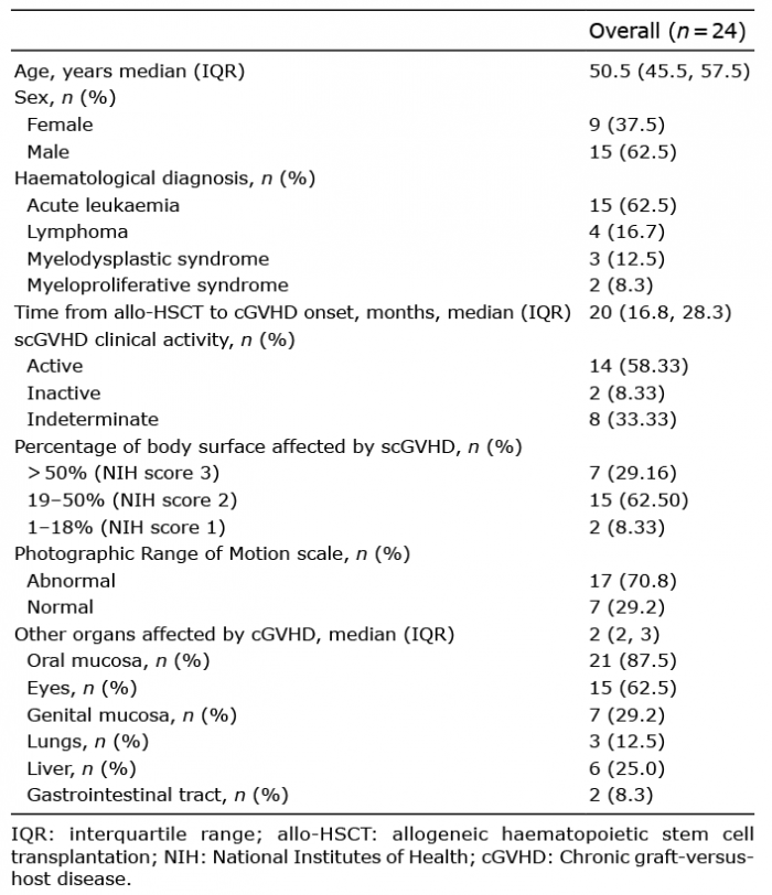 Click to show fullsize
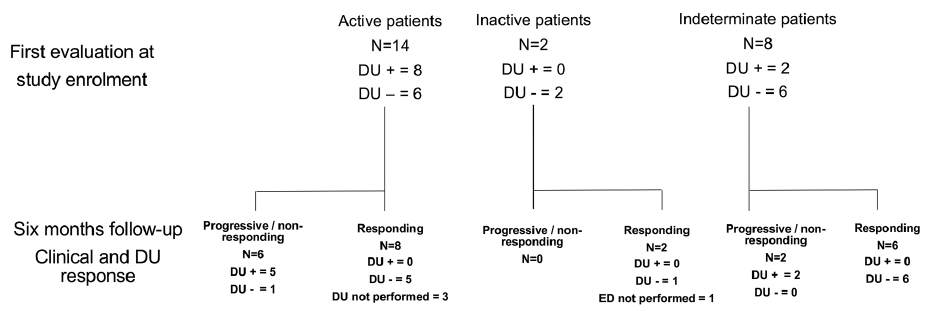 Click to show fullsize
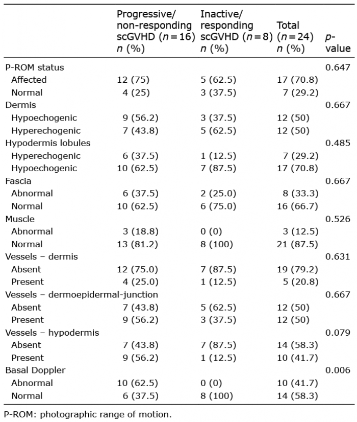 Click to show fullsize
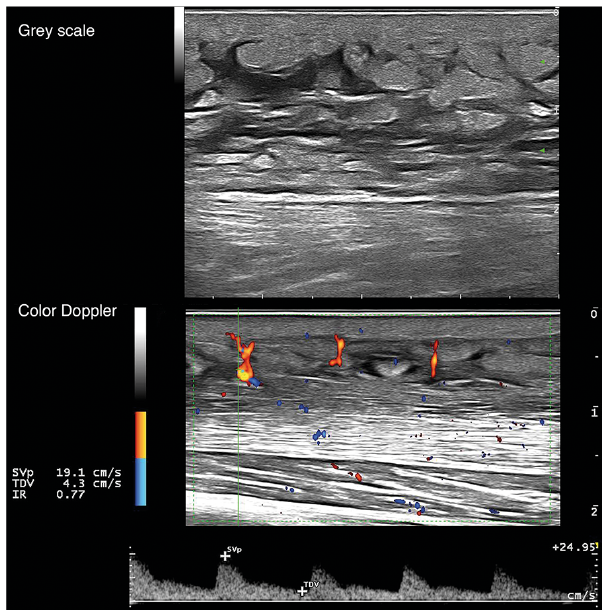 Click to show fullsize