Sir,
Musculoskeletal ultrasound (MUS) has an important role as a convenient imaging tool in the daily practice of physical and rehabilitation medicine (PRM) physicians. However, due to its major limitation, operator dependency, the optimum means of training in MUS has always been a matter of debate. Although formal direct learning provided by an expert is generally accepted as the best method, learning to perform MUS to a high standard is a challenge. It is also necessary to have a thorough knowledge of anatomy in order to comment optimally on the images; thus studying sonoanatomy has become noteworthy in this lengthy education. We describe here our experience of sonoanatomy education, in what we believe is the first presentation on this subject to the European PRM Society.
Within the course schedule of the Turkish Musculoskeletal Ultrasonography Study Group (TURK-MUSKULUS; www.turk-musclus.org) we recently organized a course entitled “AnatoMUS–I: Ultrasonographic Imaging of the Upper Limb Peripheral Nerves”. This two-day course included half a day dedicated to sonoanatomy of each of the upper limb nerves (i.e. median, ulnar and radial nerves). First, an anatomist presented a lecture on the normal anatomy of the nerve being studied. Secondly, a PRM physician presented a lecture on clinical, electrophysiological and ultrasonographical findings as regards its pertinent pathologies. Thereafter, participants studied the nerves, initially on the cadaveric specimens, then, using ultrasound, on normal live subjects (Fig. 1). At the end of each nerve session, relevant pathologies (from patient archives) were discussed as a lecture and, in the last half day, as live demonstrations on real patients.
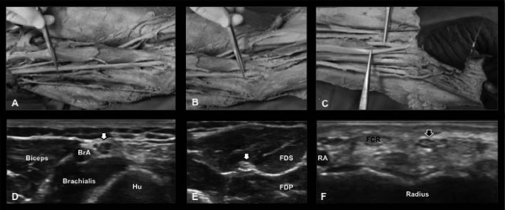
Fig. 1. Anatomical dissections of the upper limb, whereby the tweezer demonstrate the median nerve at the level of the (A) arm, (B) elbow and (C) wrist (the second tweezer on the ulnar side holding the flexor carpi radialis tendon). Ultrasonographic images (axial view) illustrate the median nerve (arrows) taken at the level of the (D) arm, (E) forearm and (F) wrist. The fascicular pattern of the nerve is visualized next to the brachial artery (BrA) while passing close to the humerus (Hu). In the forearm, the nerve courses between the flexor digitorum superficialis (FDS) and flexor digitorum profundus (FDP) muscles. Distally, the nerve is localized quite superficially ulnar to the flexor carpi radialis (FCR) tendon and the radial artery (RA).
Depending on the availability of (in particular fresh-frozen) cadavers, MUS courses using anatomy specimens indisputably provide great opportunities to study sonoanatomy of the musculoskeletal system, for both diagnostic and therapeutic interventions alike. In AnatoMUS–I we chose to start with the peripheral nerves, because MUS is very convenient for scanning a peripheral nerve from its proximal plexus to its distal small branches (e.g. for the median nerve, from the brachial plexus to its palmar branches). Furthermore, PRM physicians commonly deal with peripheral nerve pathologies for which, in addition to electrodiagnostic tests, MUS can reveal 3 important aspects (1). For example, in the case of peripheral nerve entrapment syndromes, first it can morphologically confirm the presence of pathology (i.e. nerve swelling), secondly it can uncover the underlying cause (e.g. an adjacent ganglion cyst or a bony spur), and thirdly it can provide guidance during onward interventions (e.g. aspiration or injection) (2).
In conclusion, it is hoped that our experience will facilitate future educational development in this expanding PRM discipline. AnatoMUS–II, for the lower limb peripheral nerves, has already been scheduled, and the next stage of progress could be achieved by the use of fresh cadavers simultaneously to scan and dissect the structures under investigation.
References
Submitted February 20, 2012; accepted March 1, 2012.
Levent Özçakar, MD1*, Bülent Yalçın, MD2, Murat Kara, MD3, Elif Yalçın, MD3, Tülay Tiftik, MD3, Seda Gülbar, MD4, Sedat Develi, MD2 and Fatih Yazar, MD2
From the Departments of 1Physical Medicine and Rehabilitation and 4Anatomy, Hacettepe University Medical School, 2Department of Anatomy, Gülhane Military Medical School and 3Ankara Physical Medicine and Rehabilitation Education and Research Hospital, Ankara, Turkey. *E-mail: lozcakar@yahoo.com

