


Maria Claudia Tirico, Cyro Festa Neto, Neusa Yuriko Sakai Valente and Marcello Menta Simonsen Nico*
Department of Dermatology, University of São Paulo, Rua Itapeva 500-3A, 01332-000 São Paulo, SP, Brasil. E-mail: mentanico@hotmail.com
Accepted Aug 25, 2015; Epub ahead of print Aug 28, 2015
Although the injection of paraffin, mineral oil, and silicon into body parts began to be described in the 19th century, similar procedures had been practiced before that time (1). Women undergo wrinkle correction and breast augmentation, whereas men seek to achieve erectile enhancement and penile enlargement (2–5). Reports on the development of subcutaneous nodules at the sites of injection abound – commonly referred to as sclerosing lipogranuloma (SL) and paraffinoma (6).
SL and paraffinoma are types of foreign body panniculitis that are characterized histopathologically by fat globules that vary in size, replacing subcutaneous fat tissue. These bodies are surrounded by multinucleated foreign body giant cells and are associated with chronic granulomatous inflammatory reactions, free fat droplets, hyalinization of fibrous tissue, fibrosis; calcification can occur in long lasting lesions (6).
Visceral lesions of LG are rare and can lead to death (7, 8). We report an unusual case of SL with widespread cutaneous lesions and pulmonary compromise.
case Report
A 57-year-old married man presented with painful cutaneous enlargements that had persisted for 10 years. He was a construction worker who was retired from his job because of his skin disease.
The examination showed several nodules that converged into firm bulky masses in the genital area, including the penis and scrotum, extending to the suprapubic region. The anterior and posterior neck areas showed voluminous and deforming masses, and the right forearm and left hand were also affected (Fig. 1). The lesions had an irregular surface and were hard on palpation; pain was relieved only with a combination of opioids and antidepressants.
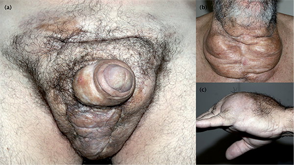
Fig. 1. (a) Voluminous genital mass extending to the supra pubic area, (b) Converging nodules on the anterior cervical region, (c) Soft tissue enlargement of the dorsum of the left hand.
Since the initial presentation, the patient had been examined in many hospitals, in which he underwent several resections and received corticosteroids and antibiotics, without any response.
The histopathological aspects of several biopsied nodules were similar and included the presence of vacuoles with multinucleated foreign body giant cells, foamy macrophages, sclerosis of the dermis, and mild inflammatory infiltration of lymphocytes and histiocytes (Fig. S11). Stains for bacteria, fungi, and acid-fast bacilli and cultures were negative.
Computerized tomography scans revealed thickening and densification of the skin and subcutaneous tissue, with diffuse hyperattenuating linear foci that infiltrated the subcutaneous tissue, corresponding to exogenous substances or calcification. Multiple hypoattenuating signs were interpreted to have resulted from previous inflammation and the presence of gas that permeated the tissues. Nodules and tumors, up to 5 cm in diameter and having similar characteristics, were detected in the lung parenchyma bilaterally, associated with a high number of thoracic lymph nodes (Fig. S21). Bronchoscopy with lung biopsy was then performed, which revealed a foreign body granulomatous reaction.
The patient, despite experiencing pain, proudly exhibited his enlarged genitals but firmly denied injecting any substances into the affected sites, blaming medical malpractice for his prolonged illness. The patient and his family had a troubled relationship – his wife referred to him as “intensely jealous,” and he had attempted suicide several years earlier. He demanded constant attention from his family (wife and 4 adult daughters) concerning his disease.
After refusing for several weeks, the patient finally agreed to be examined by a psychiatrist. In the consultation, the patient insisted on almost solely discussing the purely cutaneous aspects of his disease, failing to provide details about his mental health. The psychiatrist’s clinical impression was that the patient had personality disorder with features of manipulative personality and factitious disorder.
The patient refused to undergo any further psychiatric evaluations.
Discussion
The differential diagnosis of panniculitides is extensive, with many causes (9).
Factitial, or foreign body, panniculitis is a specific diagnosis with various clinical findings, depending on the inciting agent; on histopathology, it usually presents as acute lobular panniculitis that is associated with fat necrosis and large inflammatory infiltrates, comprising primarily neutrophils (6).
Sclerosing lipogranuloma (SL) is a distinct type of factitial panniculitis in which chronic findings prevail- aspects include foreign body giant cell reactions, foamy macrophages, sclerosis of the dermis, and inflammatory infiltrate with lymphocytes and histiocytes. Calcification can occur in long-standing cases (3–6).
Despite the firm denial and lack of confirmation of the nature of the injected material by our patient, the diagnosis of SL was easily established, based on his clinicopathological and psychosocial profiles.
We suspect that, beginning with injections for penile enlargement, he subsequently performed this procedure at other sites. Patients with factitial disorder typically deny their behavior due to extant personality disorder and constantly develop new lesions and seek medical care (10). These cases are difficult to treat dermatologically and psychiatrically: the lesions are usually extensive and impossible to excise, and their mental disease typically does not respond to behavioral or psychopharmacological therapy (10).
In cases like our patient, a CT scan can be a useful noninvasive diagnostic tool for determining the extent of the disease (11, 12).
A crucial facet of our case was the significant pulmonary involvement in the SL. Particles reached the pulmonary parenchyma, possibly through the blood. Few studies have examined the systemic dissemination of fat particles (7, 8, 13), and the reaction of lymph nodes and lung to mineral oil, with the typical skin lesions, has rarely been described (13). Other rarer complications include pneumonitis, alveolar hemorrhage, and respiratory failure, which might be fatal: autopsies have determined pulmonary edema to be the cause of death following penile injections (7, 8).

1http://www.medicaljournals.se/acta/content/?doi=10.2340/00015555-2229
References


