


1Department of Nuclear Medicine and 4Department of Ophthalmology, Affiliated Hospital of Inner Mongolia Medical University, Hohhot, 2Department of Radiology, the People’s Hospital of Liaoning Province, and 3Department of Dermatology, No.1 Hospital of China Medical University, 155 North Nanjing Street, Shenyang 110001, China. *E-mail: xuxuegang2749290@163.com.
Accepted Jun 14, 2018; Epub ahead of print Jun 25, 2018
Premature aging syndrome, Penttinen type (Penttinen syndrome, OMIM: 601812), is a rare progeroid syndrome characterized by a prematurely aged appearance, acro-osteolysis, loss of subcutaneous fat, translucent skin with keloid-like lesions, and other symptoms (1–3). We enrolled one Chinese patient with the characteristic presentations of Penttinen syndrome and identified a PDGFRB mutation in this patient.
The patient was an 18-year-old male who was born with a generally normal appearance. At 2 years, he was noted to have frequent micturition and an open anterior fontanel. A cranial CT scan indicated hydrocephalus. At about 4 or 5 years, he presented with a large anterior fontanel and flat occiput, broad thumbs and halluces, developmental delay, and limited range of motion of the fingers. Gradually, his skin became very thin, dry, and translucent with diffuse hyperpigmentation and scattered keloid-like nodular lesions after wounds and on the pressures sites. At 18 years, he was referred to our department owing to his specific facial features. He was 192 cm tall and weighed 87.5 kg. He had marked maxillary retraction, pseudoprognathism, proptosis, ectropion, an open anterior fontanel, and a flat occiput (Fig. 1a, b). The eruption of his permanent teeth was delayed, and some deciduous teeth were still present. Dermatologic examination showed thin, dry, pruritic skin, diffuse hyperpigmentation, and loss of subcutaneous fat. His skin was particularly translucent, with a prominent venous pattern (Fig. 1c). Other findings included keloid-like nodules on the elbows, knees, and other pressure points; cicatricial contractures on the palms; and several hypopigmented lesions on the trunk (Fig. 1d–f). A biopsy of the skin on his leg showed increased melanin in the basal layer of the epidermis, homogeneous collagen, and a reduced number of sweat glands and other appendages (Fig. 1h).
Skeletal examination showed severe contractures and shortening of his fingers (Fig. 1d). He also presented with stiffness of his hands, fingers, and large articulations as well as kyphoscoliosis (Fig. 1g). X-rays confirmed these findings and showed flexion deformities of the interphalangeal joints, and acro-osteolysis of the distal phalanges, and scoliosis (Fig. S1a, b). Cranial CT and MRI scan showed thin calvarium; open anterior fontanel, posterior fontanel and sagittal suture; hydrocephalus; and temporalis hypertrophy (Fig. S1c, d). Numerous arachnoid cysts and mega cisterna magna were also noted in addition to cerebellar atrophy and leukoence-phalopathy of the bilateral periventricular white matter and centrum semiovale, which did not match to his age (Fig. S1e). Echocardiography showed left ventricular diastolic dysfunction (E/A<1). His intelligence and other examinations were found to be normal.
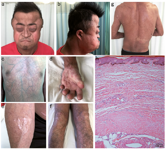
Fig. 1. (a, b) The patient had a distinctive appearance of premature aging: maxillary retraction, pseudoprognathism, proptosis, ectropion, and flat occiput. (c) Thin and translucent skin with loss of subcutaneous fat. (d) Severe contractures and shortening of the fingers. Scar-like lesions on the palm. (e) Keloid-like lesions on the elbow. (f) Diffuse hyperpigmentation and nodules on the lower extremities. (g) Kyphoscoliosis. (h) Histological presentations of a skin biopsy from his leg showed increased melanin in the basal layer of the epidermis, a relatively thin dermis and little subcutaneous fat, a deposit of homogeneous material in the dermis and a reduced number of sweat glands and other appendages (hematoxylin and eosin, ×100). Written permission is given by the patient to publish these photos.
To identify the causative mutation, whole exome sequencing was performed as reported (4). The Institutional Review Board and the Ethics Committee of No.1 Hospital of China Medical University approved the study. The targeted exon sequences plus ?anking sequences were speci?cally captured and enriched using an array-based hybridization chip (xGen® Exome Research Panel v1.0, Integrated DNA Technologies, CA) followed by HiSeq X10 (Illumina, San Diego, CA) sequencing. The average depth for all targeted exons was 94.49× with coverage of 99.4%. All variants were filtered against several public databases (ExAC, gnomAD, 1000 genomes project). Finally, a heterozygous variant (c.1994T>C, p.Val665Ala) was identified in exon 14 of PDGFRB (platelet-derived growth factor receptor β, GenBank: NM_002609) (Fig. S2a). This variant was detected with a sequencing depth of 34× and a ratio of 0.52, which was not identified in all other family members and confirmed to be a de novo variant via Sanger sequencing (Fig. S2b–e). Thus the diagnosis of Penttinen syndrome is determined in our patient.
Penttinen et al. (1) first described a patient presenting with a prematurely aged appearance; delayed bone maturation and dental development; pronounced acro-osteolysis with brachydactyly; and distinctive cutaneous findings, including loss of subcutaneous fat. Later Zufferey et al. (2) reported 2 cases with similar presentations. Johnston et al. (3) reported another 2 cases and identified a point mutation of PDGFRB in 4 reported cases of Penttinen syndrome. Here we report a patient with the characteristic presentations of Penttinen syndrome. Besides skin and skeletal abnormalities, he had hydrocephalus since early childhood, remarkable deformities of the ventricular system, leukoencephalopathy, and cerebellar atrophy, among other anomalies. His neurological atrophy and diastolic cardiac dysfunction did not match his age and needed close follow-up to determine whether they posed a risk of premature death.
PDGFRB is a typical tyrosine kinase receptor (6). Binding of PDGFs to PDGFRB leads to the autophosphorylation of PDGFRB and subsequent activation of the downstream pathways, including mitogen-activated protein kinases (MAPKs), phospholipase Cγ (PLCγ), signal transducers and activators of transcription (STAT), and phosphatidylinositol-3 kinase (PI3K) (6, 7). Mutations in PDGFRB have been documented in a wide range of phenotypes. Loss-of-function mutations in PDGFRB lead to idiopathic basal ganglia calcification-4 (IBGC4) (8). PDGFRB mutations also cause infantile myofibromatosis (IM), a common benign fibrous tumor of soft tissues (9). Recently, germline mutations in PDGFRB have been associated with a novel overgrowth syndrome, named Kosaki overgrowth syndrome (10, 11). The PDGFRB gene has also been found to fuse with more than 36 other genes, and they are involved in many myeloid and/or lymphoid neoplasms (12).
PDGF signaling has been implicated in organ fibrosis and is assumed to play a role in driving the proliferation of cells of mesenchymal origin (7, 13). Johnston et al. (3) identified the downstream effectors of the p.Val665Ala PDGFRB variant, including STAT3 and PLCγ1. STAT3 was proved to contribute to keloid pathogenesis by promoting collagen production, cell proliferation and migration (14). Craggs et al. (15) revealed increased expression of PDGFRB in pericytes in cerebral autosomal dominant arteriopathy with subcortical infarcts and leukoence-phalopathy. Taken together, we hypothesize that excess, ligand-independent phosphorylation of PDGFRB leads to the activation of downstream signaling and causes the presentations of Penttinen syndrome.
We thank the patient and his family. This work was supported by National Natural Science Funding of China (code: 81602740) and the National Key Research and Development Program of China (No.2016YFC0901504).
The authors have no conflict of interest to declare.


 Click to show fullsize