


Department of Dermatology, The First Hospital of China Medical University and Key Laboratory of Immunodermatology, Ministry of Health and Ministry of Education, Nanjing Street, Shenyang 110001, China. *E-mail: lizhang_1001@126.com
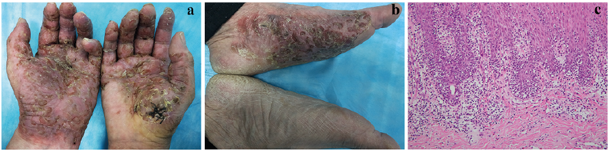
A 53-year-old man was referred with a 7-year history of erythematous patches and plaques containing scales and fissures. This condition had been present on both palms and the left sole, and, over the past 2 years, it had progressed to involve the nails. The eruption had been asymptomatic, except for occasional episodes of pruritus. For several years he had been diagnosed with eczema and was treated with topical corticosteroids that yielded only suboptimal improvements. Over the past 10 days, he noticed the appearance of numerous small pustules with a yellowish crust that were confined to the palms and soles. His medical and family history were unremarkable.
Physical examination revealed well-demarcated dark-reddish plaques with adherent crusts, occasional erosion and fissuring, involving both palms and surfaces of fingers as well as the left sole. In addition, a dystrophy and yellow discoloration was present in several finger nails (Fig. 1a, b). Fungal examination was negative and routine blood tests and imaging examinations were normal. An initial diagnosis of palmoplantar pustulosis was formulated.
A skin biopsy showed hyperkeratosis with dense infiltration of atypical lymphocytes lining the dermo-epidermal junction and partial lymphocytes infiltrating into the epidermis forming Pautrier’s microabscesses (Fig. 1c). Immunohistochemical staining revealed that intraepidermal lymphocytes were positive for CD3, CD4, but negative for CD7, CD8, and CD20.
What is your diagnosis? See next page for answer.
Fig. 1. (a, b) Clinical appearance of erythematous patches and plaques with scales and fissures. (c) Histopathology biopsy findings revealed dense atypical lymphocytes lining the dermo-epidermal junction and partial lymphocytes escaping into the epidermis to form Pautrier’s microabscesses (haematoxylin and eosin (H&E), ×400).
Acta Derm Venereol 2020; 100: adv00186.
Diagnosis: Mycosis fungoides palmaris et plantaris (MFPP)
T-cell receptor Jβ- and Jg-chain monoclonal gene rearrange-ments were positive, which further confirmed the diagnosis of MFPP. The patient was subsequently treated with narrow-band ultraviolet B (UVB) phototherapy at an initial dose of 0.2 J/cm2 3 times per week with gradual incremental doses, to a maximum dose of 3.6 J/cm-2. In addition, a systemic injection of interferon-α1/b at a dose of 30 μg was administered every other day, together with topical bexarotene. The skin lesions, including nail changes, regressed slowly over a 4-month period following initiation of this treatment, leaving healthy pink skin on the palmoplantar surfaces. No recurrence was evident at a 1-year follow-up (Fig. 2).
MFPP is a rare variant of cutaneous T-cell lymphoma. It presents as a wide spectrum of clinical manifestations limited to the palms and soles, including hyperkeratotic plaques, verrucous nodules, pustules, vesicles and mimics palmoplantar psoriasis, hand eczema, hyperkeratotic lichen planus, contact dermatitis, secondary syphilis, dermato-phytosis, and verruca (1, 2). Accordingly, it is quite difficult to correctly diagnose MFPP based on its clinical features, especially when there is no extracutaneous presentation and accompanying nail involvement. As MFPP does not generally have any aggressive features with local recurrences, and no lethal cases have been documented to date, it can be easily overlooked or misdiagnosed as common benign dermatoses, leading to a delay in diagnosis and treatment (3). This case indicates that mycosis fungoides may involve the palms, soles and nails, and also display asymmetrical features. Histopathology, immunohistochemistry and T-cell receptor (TCR) gene rearrangement analysis of this asymptomatic lesion may be helpful to distinguish early MFPP and recalcitrant palmoplantar dermatosis.
Fig. 2. Combination of narrow-band ultraviolet B (UVB) phototherapy and injection of interferon-α1/b markedly improved skin lesions and nail changes after 12 months.
This research is supported by grants from the National Natural Science Foundation of China (81803148).


 Click to show fullsize
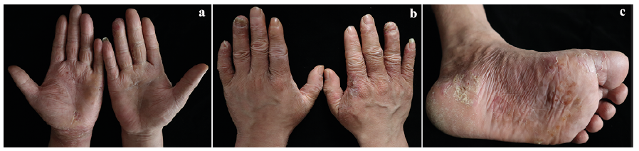 Click to show fullsize