


André Oliveira1, Iris Zalaudek2, Edith Arzberger2, Cesare Massone2 and Rainer Hofmann-Wellenhof2
1Department of Dermatology, Hospital de Santo António dos Capuchos – Centro Hospitalar de Lisboa Central, Alameda de Santo António dos Capuchos, Lisbon, Portugal, and 2Department of Dermatology, Medical University of Graz, Graz, Austria. E-mail: andre.oliveira@sapo.pt
Accepted Mar 10, 2016; Epub ahead of print Mar 15, 2016
Mammary Paget’s disease (MPD) is an uncommon intra-epidermal adenocarcinoma of the nipple-areola complex, occurring in 1–5% of all breast carcinomas (1). MPD is difficult to diagnose clinically as it mimics a variety of both inflammatory and neoplastic skin diseases (2). Pigmented mammary Paget’s disease (PMPD) corresponds to an even less common variant, frequently simulating other pigmented lesions of the nipple, including melanoma (3–5). According to the epidermotropic theory, Paget cells (PCs) originate from cancer cells that migrate via the lactiferous ducts along the basal membrane, to invade the epidermis of the nipple and areola (6, 7). Considering its intra-epidermal spreading, PCs are therefore potentially demonstrable using non-invasive diagnostic techniques with near-cellular resolution, such as reflectance confocal microscopy (RCM) (8). The aim of this study was retrospectively to describe the RCM features of 5 cases of MPD, with dermoscopic and histopathological correlation.
MATERIALS AND METHODS
Clinical, dermoscopic and RCM images from 5 women (age range 49–83 years; mean 65 years) with a diagnosis of MPD according to histopathological features and immunohistochemical reaction pattern were retrospectively collected from the database of the Department of Dermatology, Medical University of Graz, Graz, Austria. Confocal images were obtained using a near-infrared, reflectance mode, confocal laser-scanning microscope (VivaScope1500®, Caliber: imaging and diagnostics, Rochester, NY, USA). Following dermoscopic and RCM imaging, a punch biopsy from each suspicious lesion of the nipple was performed for histopathological evaluation and immunostaining.
RESULTS
Examples of clinical and dermoscopic images are shown in Fig. 1. Typical clinical presentation of MPD was seen in all but one case (patient 5): well-demarcated eczema-like plaques involving the nipple/areola complex; erosive, oozing or fissured surface; and brown-to-yellow crusts or scales. Patient 5, a 58-year-old woman, presented with a 2-month history of a fast-growing, partially pigmented nodule on the left nipple, mimicking melanoma under clinical inspection. Histopathological examination revealed in all cases a proliferation of large PCs with hyperchromatic nuclei and abundant clear cytoplasm. These cells stained positively for cytokeratin 7 (CK7) in all cases, supporting the diagnosis of MPD. As clinicopathological correlation could not rule out melanoma in patient 5, further immunostaining for S-100 protein and HMB-45 was also performed. PCs were negative for both. This profile allowed not only the exclusion of melanoma, but also the confirmation of PMPD. Additional diagnostic procedures included breast ultrasound and mammography, which were followed by surgical excision. An underlying ductal carcinoma in situ (DCIS) of the breast was found in all patients. Dermoscopy disclosed non-specific findings in all 4 erythematous lesions: a pink-whitish to red background was common to all cases; polymorphous vessels (3/4); erosions (2/4); yellow scales (2/4); and shiny-white streaks (1/4). In patient 5, dermoscopy revealed polymorphous vessels irregularly distributed within a red-to-yellow, whitish background; chaotic spread of superficial brown dots; and structureless areas of grey pigmentation. Imaging of the normal appearing, contralateral nipple was performed in patient 1 using RCM. A lobular arrangement of normal honeycomb pattern was observed in all epidermal layers, reflecting the nipple architecture (Fig. 2a). This pattern was lost in all lesions due to prominent pagetoid spread and disarranged architecture (Fig. 2b). Poorly reflective round cells surrounded by a dark stroma were seen as dark holes, mostly at the stratum corneum (SC) and granular layer (GL) levels, corresponding to PCs (Fig. 2c). These cells were 1.5–2 times larger than close keratinocytes, which were often distorted and elongated (Fig. 2d). Single cells or small nests of cells with a bright central area and a peripheral large dark halo, appearing as target structures, were also observed at GL in all lesions (Fig 2e). Further RCM images showing particular features of MPD are shown in Fig. S2f–j1.
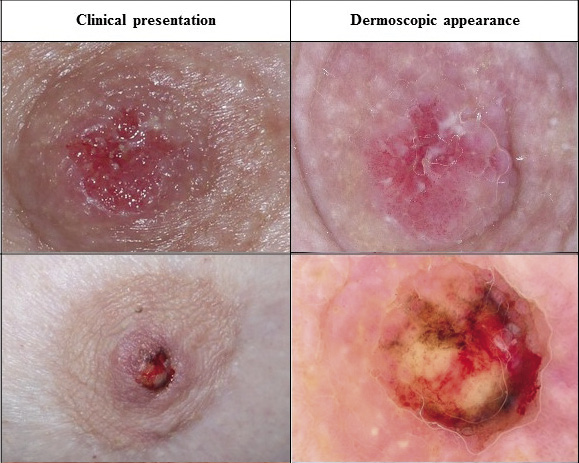
Fig. 1. Clinical and dermoscopic appearance in mammary Paget’s disease. Patient 1 (top) shows a typical appearance; patient 5 (bottom) an uncommon pigmented form. Photographs for all 5 patients are shown in Fig. S11.
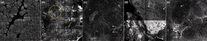
Fig. 2. Reflectance confocal microscopy: (a) typical honeycomb pattern in a normal nipple (mosaic, 6 × 6 mm). (b) Disarranged epidermal architecture (patient 5, pigmented mammary Paget’s disease); multiple tumour cells seen as isolated dark holes (white arrows) or target structures clustered in small (blue arrow) or large nests (yellow arrow); dendritic cells (yellow circle) in the upper-epidermis (mosaic, 3.5 × 3.5 mm). (c) Paget cells found at superficial epidermal levels as isolated dark holes (white arrows) or small nests (blue arrows), resembling target structures (white star) (basic image, 0.5 × 0.5 mm). (d) Paget cells (white arrows) in the stratum corneum were 1.5–2 times larger than neighbouring keratinocytes (mosaic, 2 × 2 mm). (e) Larger nests (yellow arrows) appearing both as dark holes (white arrow) or target structures (white star) observed at granular layer (basic image, 0.5 × 0.5 mm).

1http://www.medicaljournals.se/acta/content/?doi=10.2340/00015555-2402
DISCUSSION
This study confirms previous reports showing that RCM can be used to provide in vivo images of poorly reflective PCs, appearing both as dark holes or target structures (8–10). The dark halo found in target structures probably relates to clefts between PCs and the surrounding epidermis due to mucin secretion by the former. These findings are in agreement with the described glandular features of MPD in immunohistochemistry (7). Dark holes correspond to large cells with abundant and pale cytoplasm, while bright central areas in target structures relate to pleomorphic nuclei. Confocal features of PCs have an excellent correlation with the histopathological presentation of atypical cells scattered throughout the epidermis, isolated or in nests of heterogeneous sizes (1, 2). Pagetoid spread of cells 1.5–2 times larger than adjacent keratinocytes is a suggestive feature of MPD in RCM. Therefore, larger size of cells may help to differentiate MPD from other skin tumours with confocal Pagetoid spread, including melanoma, Bowen’s disease, Spitz naevus, mycosis fungoides and Bowenoid papulosis (9, 10). A higher density of nests was found in deeper layers of the epidermis, dermal–epidermal junction (DEJ) and upper dermis. Therefore, limited depth examination of the papillary dermis in RCM may prevent the identification of an underlying invasive MPD. Disarranged epidermal architecture with loss of normal honeycomb pattern was also a striking RCM feature in all studied cases. Hence, monotonous architectural and cytomorphological findings will help to differentiate MPD from common inflammatory diseases, such as atopic eczema or contact dermatitis. RCM presentation of atopic eczema and contact dermatitis may include spongiosis, exocytosis, intra-epidermal vesicles and regular, focally blurred, honeycomb pattern (11). PMPD is a rare variant that may simulate melanoma, not only clinically, but also in its dermoscopic and histopathological presentation (3, 5). RCM was not an exception to such diagnostic difficulties, because atypical, large pagetoid cells, together with a disarranged epidermis are also found in melanoma. Although immunohistochemical analysis remains the mainstay for definitive diagnosis, RCM may provide additional non-invasive clues: e.g. a higher density of tumour nests in the DEJ and superficial dermis; bright collagen bundles due to stroma reactivity to such nests; dendritic structures in the epidermis corresponding to melanocytes; roundish, bright cells at the DEJ relating to melanophages; increased vascularization within tumour nests; larger Pagetoid cells than in melanoma; and brighter when compared with the described double appearance of PCs in erythematous lesions of MPD, probably due to increased melanin content. Dermoscopy of erythematous, eczema-like MPD revealed non-specific findings, while in PMPD, superficial chaotic brown dots and grey structureless areas probably relate to epidermal melanocytes and clusters of tumour cells with increased melanin.
REFERENCES


